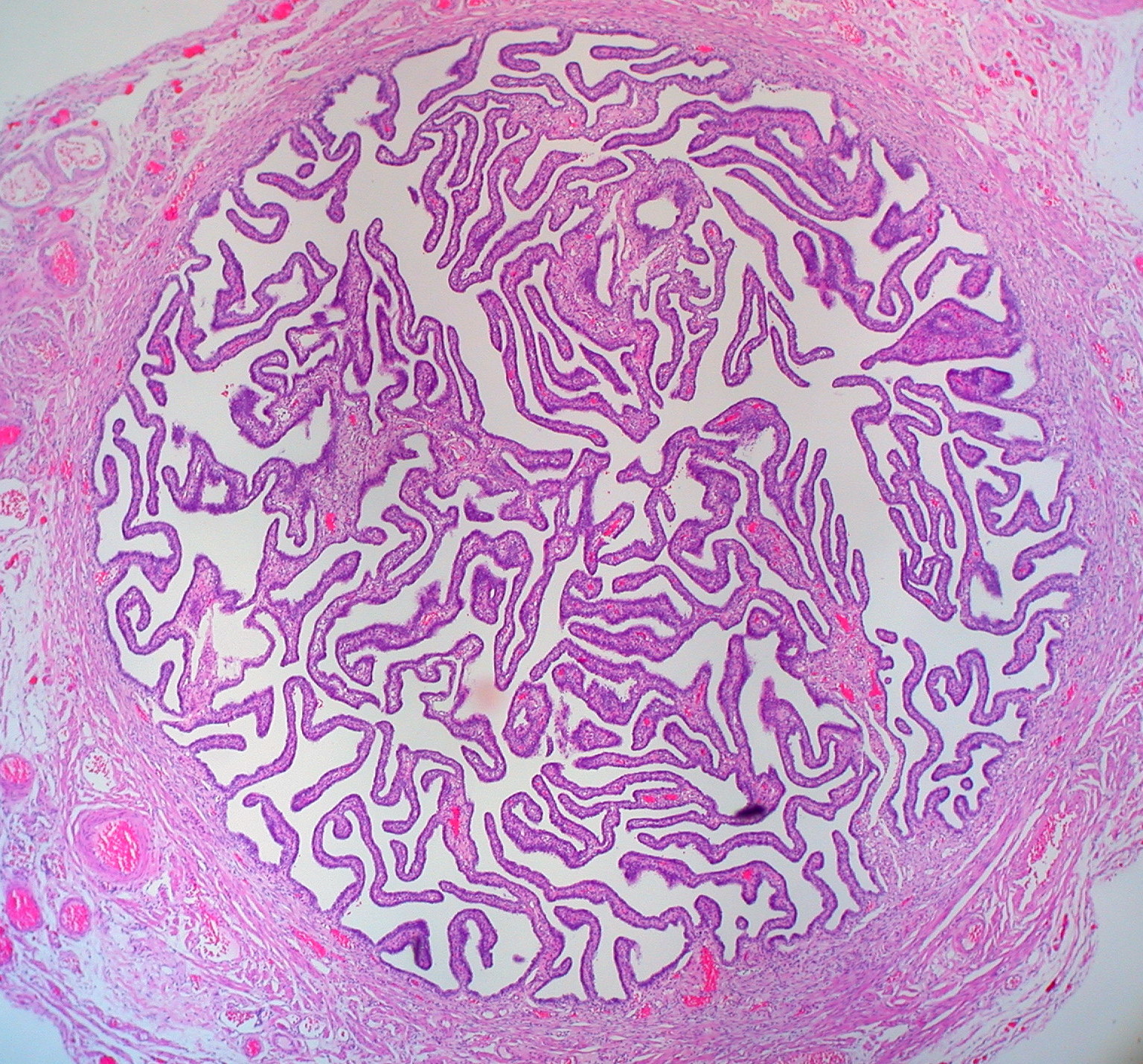
Uterine tube
The Tuba Uterina (fallopian tube, salpinx, Fallopian tube) is a hollow paired tube 8-15 cm long. It is attached to the ligamentum latum uteri (plica lata uteri). The fallopian tube hanger itself is called the 'mesosalpinx' and forms the upper part of the 'ligamentum latum uteri'. The lateral end of the fallopian tube is opened into the peritoneal cavity (ostium abdominale tubae uterinae), the medial end into the uterine cavity.
Parts of Fallopian Tube[edit | edit source]
The fallopian tube has the following parts: infundibulum, 'ampulla', 'isthmus' and 'pars uterina .
The Infundibulum is the funnel-shaped part facing the ovary. About 10 projections of various lengths - 'fimbriae', extend from its edge, the longest of which is called 'fimbria ovarica and is fixed to the surface of the ovary.
The Ampulla' is spindle-shaped and occupies 1/2–2/3 of the length of the oviduct. It is followed medially by a narrow and straight 'isthmus' (about 1/3 of the length of the fallopian tube) and after it by the about 1 cm long 'pars uterina', which pierces the wall corner of the uterus and opens into the uterine cavity as ostium uterinum tubae uterinae'.
The fallopian tube cavity is most spacious in the ampulla region (up to 1 cm in diameter) and narrowest in the pars uterina (less than 1 mm; 0.1–0.5 mm).
Position and Syntopy of Fallopian Tube[edit | edit source]
From the mouth to the uterus, the isthmus tubae stands transversely and points across to the wall of the small pelvis. The lateral section of the tube bends dorsocranially; the infundibulum tubae with fimbriae folds in situ dorsocaudally to the ovary and the fimbriae reach the upper medial side of the ovary. The fallopian tubes are in contact with the loops of the small intestine. The right fallopian tube lies close to the appendix, the left is in contact with the colon sigmoid.
Oviduct Construction[edit | edit source]
 The wall of the fallopian tube consists of three layers - mucosa, smooth muscle and peritoneum.
- Tunica mucosa - composed of cilia - they are highest and most numerous in the ampulla, where they form an articulated labyrinth also composed of secondary and tertiary cilia. It is covered by a single-layered cylindrical epithelium with ciliated and secretory cells, it also contains undifferentiated cells from which the epithelium regenerates. The Lamina propria is made up of a sparse collagenous tissue. Algae and ciliated cells decrease from the periphery to the uterus, while secretory cells increase in the same direction.
- Tunica muscularis - from the periphery to the uterus it is thicker, but it is the thickest in the isthmus, it consists of the outer longitudinal layer and the inner circular layer.
- Tunica serosa -> it is an intraperitoneal organ
Function of fallopian tube[edit | edit source]
Just before ovulation, the infundibulum moves along the surface of the ovary with active smooth muscle contractions to the places where the mature follicle is located. After ovulation, the released egg is sucked into the fallopian tube cavity. Fertilization usually takes place in the ampoule, and the fertilized egg is transported to the uterine cavity during the next 4-5 days by the movement of the cilia and the peristalsis of the tube. An unfertilized egg undergoes autolysis. The tubal muscle also has the opposite effect - an antiperistaltic movement that helps sperm penetrate to the egg.
Vascular supply and innervation[edit | edit source]
- Arteries: anastomosis r. tubarius a. uterinae and r. tubarius a. ovaricae.
- Veins: v. ovarica, plexus venosus uterovaginalis.
- Lymph: nodi lymphatici lumbales.
- Nerves: plexus ovaricus, plexus uterovaginalis, mainly autonomic.

