Tibial nerve palsy
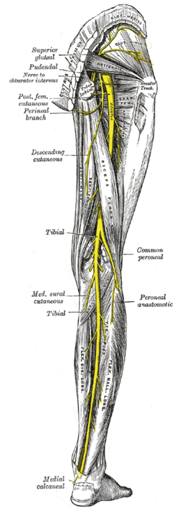
The Nervus tibialis is formed by fibers from the L5–S2 roots. It separates from n. ischiadicus, sends several motor branches (m. triceps surae, m. tibialis post., m. flexor digitorum longus and flexor hallucis longus) and sensitively innervates the back of the calf and the lateral part of the leg (creates a connection with the peroneus nerve and creates a sensitive suralis nerve). It passes through a strait at the tarsal tunnel (behind the inner ankle, covered by the retinaculum of the flexors) and in the porta pedis (the course of the nerve or its final branches - the nn. plantares in plant pod m. abductor hallucis). During the course, the vulnerable place is in the fossa poplitea (nerve located superficially in the axis of the limb).
Image of polio[edit | edit source]
- it is impossible to stand on tiptoes, lift the heel - when walking, the patient stomps with his heels
- sensitivity disorders in the innervation area (mainly planta)
- the Achilles tendon reflex often disappears
Causes[edit | edit source]
Individual disability is very rare. Compared to the "n. peroneus", it is significantly less fragile.
- knee trauma — dislocations and displaced fractures
- trauma in the area of the passage behind the inner ankle — cuts and lacerations, ankle fractures, pressure with a plaster cast, etc.
- syndrome tarsal tunnel — initially manifested as intermittent pain shooting into the plantar, with a longer duration of constant paresthesia and pain
Links[edit | edit source]
Related Articles[edit | edit source]
- Tibial Nerve
- Ischiadicus
- Tarsal Tunnel
- Fossa poplitea
- [[Peripheral nerve involvement syndrome
Source[edit | edit source]
- AMBLER, Zdeněk – BEDNAŘÍK, Josef, et al. Klinická neurologie. 1. edition. Praha : Triton, 2010. ISBN 978-80-7387-389-9.

