Syphilis (clinical picture)
Acquired syphilis typically develops in three clinical stages.
Primary stage[edit | edit source]
The primary stage is characterized by changes in the site of infection. The incubation period from infection to manifestations on the mucosa or skin is about 21 days. The following lesions are characteristic of the primary stage of syphilis:
- Ulcus durum (hard ulcer):
- at first a flat papule changing gradually in erosion and ulcer;
- raised margins, deep red base;
- palpable (gloved!) stiff ulcer with indurated edges;
- painless;
- lymphocytes, plasma cells and macrophages predominate in the infiltrate;
- treponems survive intracellularly after the primary infection has healed.
2. Regional lymphadenitis (swelling of the descending nodes)
- arises in 1-2 weeks;
- the nodules are enlarged, painless and reddish.
Antibodies generated in the primary stage do not prevent the development of the secondary stage.
Secondary stage[edit | edit source]
The secondary stage is a manifestation of the generalization of the infection, it occurs within two years of the primary infection. Treponemata multiply in the bloodstream. The manifestation is maculopapular rash on the skin and mucous membranes. The concentration of antibodies increases significantly and immunocomplexes are formed.
- Manifestations of generalization of infection
- skin rash - macules or papules 0.5–1 cm, red, may be covered with scales
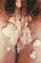
- condylomata lata - papules to bumps, red or skin color;
- leucoderma syphiliticum - depigmentation of the neck;
- on the mucous membranes of the enanthema (mucous membranes of the mouth, genitals);
- alopecia areolaris (diffuse alopecia in the forehead and beard, loss of eyelashes and lateral thirds of the eyebrows);
- hepatitis, lymphadenitis (lasts 2-8 weeks), from 6 months in 25% neurological symptoms - acute syphilitic meningitis.
The first and second stages are contagious.
Tertiary stage[edit | edit source]
The tertiary stage is a late and non-infectious manifestation of lues after varying lengths of latency. Treponemata show up only rarely. Organ changes develop, vascular damage and CNS damage occur.
- Manifestations of tertiary syphilis
- rubber:
- sharply demarcated bumps (on the skin and internal organs), obviously red, palpably stiff, collapsed in the center;
- the contents perforate on the outside, an ulcer is formed and a viscous yellowish fluid flows out;
- heals with a depigmented scar;
- skin - syphilis noduloulcerosa:
- papules and bumps merging into deposits of deep red color, heal in the center with an atrophic pigment scar;
- on extensors, back and face;
- in 7% neurological symptoms - progressive paralysis, dorsal tabes
- In 25% of untreated lues, acute syphilitic meningitis occurs no earlier than 6 months after primary infection;
- it is caused by the meningeal invasion of Treponema pallidum (a spirochete sensitive to environmental influences);
- does not affect the brain parenchyma.
- Some untreated obliterative endarteritis (syphilis meningovasculosa), chronic subpilar meningitis (syphilis spinalis), and optic atrophy appear 5-15 years after infection.
- Some people do not have to go through all stages of the disease, about a third of people recover after the primary infection without treatment, and another third remain infected with no clinical manifestations. Only one third of patients develop the tertiary stage.
Congenital syphilis[edit | edit source]
See the Congenital Syphilis page for more information.
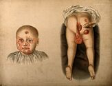
Congenital syphilis is caused by transplacental transmission of the disease from mother to fetus. In maternal infection in II. In the first trimester of pregnancy, syphilis congenita tarda (school-age manifestations, Hutchinson's Triassic and bone involvement) develops when the mother becomes infected in III. during the trimester of pregnancy, syphilis congenita praecox (manifestation in neonatal age, symptoms of stage 2 syphilis) occurs.
Hutchinson's Triassic: blindness, deafness, barrel-shaped teeth.
Links[edit | edit source]
Related Articles[edit | edit source]
- Syphilis
- Congenital syphilis
- Treponema pallidum
External links[edit | edit source]
- Syfilis (česká wikipedie)
- Syphilis (anglická wikipedie)
Reference[edit | edit source]
- ↑ SEIDL, Zdeněk and Jiří OBENBERGER. Neurology for study and practice. 1st edition. Prague: Grada Publishing, 2004. ISBN 80-247-0623-7.↑ DOSTÁL, Václav, et al. Infectious diseases. 1st edition. Prague: Karolinum, 2005. ISBN 80-246-0749-2.
References[edit | edit source]
- SEIDL, Zdeněk and Jiří OBENBERGER. Neurology for study and practice. 1st edition. Prague: Grada Publishing, 2004. ISBN 80-247-0623-7.
- POVÝŠIL, Ctibor and Ivo ŠTEINER, et al. Special pathology. 2nd edition. Prague: Galén-Karolinum, 2007. pp. 297-299. ISBN 978-80-7262-494-2.
- DOSTAL, Vaclav, et al. Infectious diseases. 1st edition. Prague: Karolinum, 2005. ISBN 80-246-0749-2.
- BEDNÁŘ, Marek, et al. Medical microbiology. 1st edition. Prague: Marvil, 1996. 558 pp. 186.


