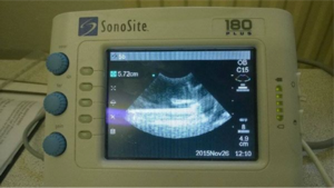
Sonography Practical
Introduction[edit | edit source]
What is Sonography?[edit | edit source]
Sonography is a painless non-invasive medical procedure that uses high-frequency sound waves to produce visual images of organs and tissues (B-mode Sonography) or blood flow (Doppler Sonography) inside the body. Depending on the situation, sonography may be used to examine the abdomen, genitalia, heart and capillaries etc. Sonography is when a sound wave strikes an object and it gets partially reflected and by measuring these echo waves, it is possible to determine how far away the object is, as well as the object’s size. As compared to other imaging techniques for viewing the internal organs like x-rays, MRIs or CT scans, the ultrasonography technique has lesser risk as it is non-ionising (compare CT which is ionising) and does not involve very strong magnetic fields (as MRI), moreover it is a very good diagnostic tool.
Brief Explanation[edit | edit source]
Sonography is based on ultrasound (frequency above 20 KHz). When high frequency sound waves travel forwards, they continue to move until they make contact with an object. Then a certain amount of the sound is reflected back. For example, sound waves go through areas that are air-filled such as the lungs these areas appear black on the screen because all sound waves are reflected at their surface and nothing passes through them – in fact they produce a vertical shadow under them. Areas filled with tissue allow some penetration and reflection of sound and produce a grayish-white image. Really hard structures such as bone produce a bright white image as the sound waves completely bounce back to the transducer. Fluid filled structures like the bladder and blood vessels also appear black because there are relatively few cells for the sound to be reflected from. A layer of aqueous gel is applied over the skin to make sure that the sound has an air-free path to the organ.The ultrasound waves penetrate the body, strike the organ moving through various types of tissue with different acoustic impedances and reflect back to the surface.The reflected waves are processed by the computer into a 2D/3D image that appears on screen. The image is called as Sonogram.
Theoretical part[edit | edit source]
Ultrasound and its properties[edit | edit source]
Ultrasound is a mechanical longitudinal sound wave with frequencies higher than the upper audible limit of human hearing (f = 20 kHz), so humans cannot hear it. It’s wavelength is very short. Ultrasound has a strong reflection at tissue interfaces. It is better absorbed by solid materials than liquids. Ultrasound travels at different speed through mediums of different materials. The frequencies used in Sonography are 2 - 15 MHz.
Acoustic impedance[edit | edit source]
Acoustic impedance is a physical quantity characterizing the acoustic property of a medium. It is defined as Acoustic impedance of a tissue = (Density of tissue) * (Velocity of sound in the tissue). At the interface of two different mediums with different acoustic impedances part of the wave is reflected and part is transmitted to the second medium. The fraction R of intensity reflected at the interface of two mediums of acoustic impedance Z1 (before reaching the interface) and Z2 (after the interface) is:
R = ((Z1 - Z2) / (Z1 + Z2))²
Between the ultrasonic transducer and the surface of the body an air layer is formed. The impedance of the air is very low compared to that of skin so R would be very large at the skin (almost 100% reflection). This means that no ultrasound would enter the body and no image is produced! To avoid this problem it is necessary to make sure that the wave travels through a medium that has similar acoustic impedance as the skin and not air, therefore during the examination a gel is applied to the area of contact of the transducer with the skin.
Ultrasound imaging[edit | edit source]
Waves that are reflected are detected using an ultrasound sensor (also called transducer) which is based on the piezoelectric effect. The waves make the crystals in the transducer vibrate, so they generate electric current that is then processed by the device. The time interval from which the ultrasound pulse leaves the transducer to when the reflected pulse is detected will provide information about the depth of the interface below the skin.
Types of areas in ultrasound image[edit | edit source]
hyperechoic - white areas on the screen (areas with high intensity of reflections)
hypoechoic - grey areas on the screen (low intensity of reflections)
anechoic - black areas on the screen (no reflections)
Bioeffects of ultrasound[edit | edit source]
Thermal effects (conversion of ultrasound energy to heat)[edit | edit source]
When traveling through living tissue, ultrasound causes warming of the tissue as the result of absorption of energy. The extent to which ultrasound raises the temperature of the tissue depends on the type of the tissue. The least warmed are liquids, then soft tissues. Solid tissues such as bones are the most warmed ones. Warming also depends on the length of the exposure of ultrasound, intensity of the ultrasound beam and whether the transducer held on one place or is moved around. It especially happens at the tissue interface but also in homogeneous tissue. The extent of absorption depends on the frequency of ultrasound. The higher the frequency is, the higher is the absorption and the penetration of ultrasound therefore decreases. The quality of the detail improves with increasing frequency, but the depth of the image decreases. Penetration increases with a lower frequency. In practice one can use probes with different requencies (lower frequencies are used when observing structures in depth, higher frequencies are used for structures situated closer to the the surface).
Mechanical effects[edit | edit source]
As a result of the fast compression and rarefaction of the medium as the wave passes cells and biomolecules can be accelerated which could occasionally lead to mechanical damage to the structures. When bubbes of gas are present the bubbles rapidly increase and decrease in volume - in certain cases leading to cavitation in which the bubbles are destroyed but leading to points of high pressure and temperature. These can damage the tissues.
Physico-chemical effects[edit | edit source]
Ultrasonic effects can also accelerate chemical reactions and excitation of molecules, blood circulation or metabolism in structures.
Importance in Clinical Medicine [edit | edit source]
Ultrasound examinations can help to diagnose a variety of conditions and to assess organ damage following illness.
Ultrasound is used to help physicians evaluate symptoms such as: [edit | edit source]
- Pain
- Swelling
- Infection
Ultrasound is a useful way of examining many of the body's internal organs, including but not limited to the: [edit | edit source]
Heart and blood vessels, including the abdominal aorta and its major branches, liver, Gallbladder, Spleen, Pancreas, Kidneys, Bladder, Uterus, ovaries, and unborn child (fetus) in pregnant patients, Eyes, Thyroid and parathyroid glands, scrotum (testicles), Brain, Hips and spine in infants
Ultrasound is also used to: [edit | edit source]
- Guide procedures such as needle biopsies, in which needles are used to sample cells from an abnormal area for laboratory testing.
- Image the breasts and guide biopsy of breast cancer.
- Diagnose a variety of heart conditions, including valve problems and congestive heart failure, and to assess damage after a heart attack. Ultrasound of the heart is commonly called an “echocardiogram”.
Doppler ultrasound images can help the physician to see and evaluate: [edit | edit source]
- Blockages to blood flow (such as clots)
- Narrowing of vessels
- Tumors and congenital vascular malformations
- Less than normal or absent blood flow to various organs
- Greater than normal blood flow to different areas which is sometimes seen in infections
With knowledge about the speed and volume of blood flow gained from a Doppler ultrasound image, the physician can often determine whether a patient is a good candidate for a procedure like angioplasty.
Advantages: [edit | edit source]
- Noninvasive examination, doesn’t need needles or any injections.
- Not painful.
- Widely available, easy to use and less expensive than any other imaging tests.
- Extremely safe, and doesn’t use any ionizing radiation like CT or strong magnetic fields like MRI.
- Gives clear picture of soft tissues which can’t be seen well by x-ray images.
- The preferred imaging for the diagnosis and monitoring of pregnant women, and unborn babies.
- Provides real-time imaging.
Disadvantages: [edit | edit source]
- Can't penetrate bone or gas.
- If the body size of the patient is too great, this will effect the imaging quality negatively.
- The patients physique affects the image quality. Fatty patients subcutaneous fat can degrade image quality, while leaner patients will get better results.
- Completely depends on the operator's/sonographer's expertise.
Ethics: [edit | edit source]
- In case of the fetal sex, disclosure can be done only with an adequate consent process.
- In obstetric sonography, the procedure can be performed only after 18 weeks and sessions must not exceed the limit prescribed in the guidelines.
- There must be doctor-patient confidentiality.
Risks [edit | edit source]
For standard diagnostic examination, there are no known harmful effects on humans.
Practical part[edit | edit source]
Task 1: Image of phantom structures
Record and log (or print) the image of the phantom structures in the best possible quality from different perspectives.
Task 2: Identification of internal phantom structures
Identify the individual internal phantom structures and measure their dimensions.
- Ultrasonography (Breast model template)
For the purposes of the practice, we divide phantoms (identified structures) into 2 groups:
Cysts - typically anechoic (dark, to black), often rounded, smooth, regular surface
Tumors - Typically echogenic formations (appear light, white), various, irregular shapes, with rough surface with bumps
Further classification of findings requires many other parameters and data and is therefore beyond practical exercises.
Task 3: Identification of various objects in the tray through an opaque cover.
Identify and describe the individual objects placed in the opaque tray.
Record uncleaned objects, describe their dimensions, echogenicity and load the completed template from MOODL into the report.
- Ultrasonography (Laboratory dish template)
Device description[edit | edit source]
For this particular examination we use the SonoSite 180 plus. It is a portable, software-driven and fully digitized ultrasound device. The system also allows ECG measurement, biopsy and any manipulation of captured images. In addition, downloaded data can be viewed on the monitor, transferred to a personal computer, or subsequently processed.
SonoSite 180 plus controls
1. Power switch
2. Near - near field gain
3. Far - far field gain
4. Gain - Total gain controller
5. Gain menu
6. Optimization, Depth and Magnification
7. Control ball
8. Patient - device setup menu
9. Function button
10. LCD screen brightness control
11. Battery charge indicator
12. LCD Contrast Controller
13. LCD screen
14. Arrows search in the last image loop
15. Buttons to select display mode
Important Notes: [edit | edit source]
- For SAFETY reasons, the device should only be used for sonographic imaging on the provided phantom and NOT ON HUMANS.
- Accompanying the sonography device is a convex sector cardiological probe with a mean operating frequency of 2 MHz. The shape of the probe allows for a fan shaped image, where it will be narrow when close to the probe and wider as depth increases. The lower frequency of the probe allows for improved imaging of deeper internal structures. However, the probe does not allow for high quality imaging for more superficial structures.
Workflow[edit | edit source]
The device is operated manually. In a normal procedure, the treated object is treated with a surface-applied gel. If gel is not available, ordinary water is often sufficient to eliminate unwanted reflections. (TIP! Be methodical when looking for structures, for example, from top to bottom as it's easier to navigate the transducer)
1. Turn on the ultrasound device by pressing and holding the power switch on the back left side of the instrument handle, then the device will beep and the screen will light up. Before working on the task, make sure that there are no images of previous practices in the device, and if so, delete them.
2. Create a new profile for the patient on the device. Press the patient and select exam / patient information in the menu that appears next. Other options will appear, select the new patient option (at the same time deleting the previous patient data). A menu with the labels name, id, accession, exam, LMP or HR will pop up. Write the group number as the name, you do not have to fill in the other information. Pressing Patient returns you to the normal view.
3. Get to know the device, its controls, and usage options. The head of the transducer you are examining the phantom with needs to be correctly rotated or the image will be inverted laterally. Also, set the correct depth on your device from which you are examining. Use the Depth button. Note that you will need to save, transfer to the PC, print or draw the relevant images you capture and measure (the procedure for transferring, viewing, and printing images will be described below).
4. Record a distinct and clear picture of the internal structures of the phantom. Capture an image of the structure from different, ideally perpendicular, angles. Then sketch it and describe it in the log.
5. Measure the dimensions of each identified internal structure. Distance measurement is done using a control ball that moves the cursor to the selected locations. Use the freeze button to "freeze" the image when a structure of interest is in view. You will see two colorful cursors (blue and green) on the screen that can be moved using the control balls to different locations on the screen (say the left and right edge of the structure the length of which you want to measure). After the first cursor is fixed using the Select button, the second cursor is released. Fix the second cursor in the desired position and press the Select button again. The measured value is displayed at the top left of the screen. Measure the actual dimensions of the structure. Write the results in the log and compare to each other.
6.Transfer images to your computer. To do this, use the Site Link Image Manager program. In the Sono Praktikum folder (folder 'Documents' from the desktop shortcut), in create a new folder with the name of your group. Set the image saving to your folder in Site Link Image Manager. Choose Configure in the top menu bar, select Image Files Location, set Documents and settings \ USER \ Documents \ Sono Practitioner \ Your Circle Components \ Your Group Folder and Confirm. To start the transfer, click Start. The Image upload complete message appears when the transfer is done.
7. Viewing files. You may use any graphics software on your computer to view the files. You should find the pictures in the folder you created in the Patient Name.No and Exam Date.YYYY Month DD.
8. Print pictures. To do so, use Picture and Fax Viewer. Once you have opened the image in the program, click on the print icon and click Next, then mark all the images you print and confirm. Confirm the printer option and then print 4 images per page. Click Finish.
9. Delete pictures from the device.
Conclusion [edit | edit source]
In conclusion, Ultrasonography is considered one of the best non-invasive techniques used to locate objects within the body. In medicine they use this device to detect changes in shape, size and texture of organs, vessels and tissues.
The Future of Ultrasonography[edit | edit source]
In the first-generation units, image conversion was carried out by inbuilt hardware, thereby making the systems quite bulky. The current generation of ultrasound equipment uses external computing systems that perform the image construction and display. This has resulted in device portability and enhanced the computing power with major improvements in image quality. Developments in signal multiplexing technology have enabled the adoption of a new generation of 3-D and 4-D (3D and time) imaging systems. It is expected that with these factors in mind, manufacturers of 3D-US and 4D-US devices will continue their efforts toward improving calculation power.
References[edit | edit source]
- Application of Ultrasound in Medicine https://www.ncbi.nlm.nih.gov/pmc/articles/PMC3564184/
- General Ultrasound http://www.radiologyinfo.org/en/info.cfm?pg=genus
- Diagnostic ultrasound , Carol M. Rumack ... et al. 4th ed.Philadelphia, PA : Elsevier/Mosby, c2011
- Atlas d'échographie en Gynécologie - Obstétrique Auteur : Dr Aly Abbara http://www.aly-abbara.com/echographie/Atlas_echographie/atlas_echographie.html
- https://www.sonoworld.com/TheFetus/Home.aspx
- Ruth E. Moran, MD, Syed Ali MD, Spencer B. Gay, MD, William E. Brant, MD., University of Virginia Health Sciences Center, Department of Radiology https://www.med-ed.virginia.edu/courses/rad/edus/index.html
- Obstetric Ultrasound - A Comprehensive Guide to ultrasound scans during Pregnancy http://www.ob-ultrasound.net/
- Ultrasound imaging http://www.wikilectures.eu/index.php/Ultrasound_imaging
- Acoustic Impedance https://www.nde-ed.org/EducationResources/CommunityCollege/Ultrasonics/Physics/acousticimpedance.htm
- Ultrasound in various media and tissue http://www.wikilectures.eu/index.php/Ultrasound_in_various_media_and_tissues
- Ultrasound Therapy http://www.wikilectures.eu/index.php/ULTRASOUND_THERAPY


