Scintigraphy in general
It is a diagnostic method to detect γ-rays and X-rays. It uses a system consisting of a scintillation detector and a conversion-amplification system.
Construction and principle of the scintillation detector[edit | edit source]
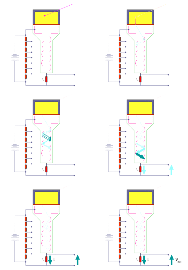
A scintillation detector consists of a scintillation crystal (usually thallium-activated sodium iodide NaI(Tl)) capable of detecting ionizing radiation in the form of a γ- or X-ray beam. The absorption of the radiation excites the electrons of the crystal and, upon their subsequent deexcitation, emits photons of visible light.[1] These very weak flashes of light are converted into a photomultiplier by suitable light guides.
The role of the photomultiplier is to multiply and transform the visible light beams into an electrical pulse consisting of a large number of electrons. This is done when light flashes from the crystal hit the photocathode. This releases a very small number of electrons from the photocathode, which interact with the dynodes (electrodes) whose surface treatment allows the pulse to be multiplied. This releases more and more electrons (on the order of 106-107 at the end), which fall as a salvo on the anode of the photomultiplier tube. This produces a measurable electrical pulse which is processed in the amplifier system.[1]
A high voltage of about 1000 V is applied between the photocathode and the anode. The environment of the photomultiplier tube is kept under vacuum.
The amplifier system consists of a preamplifier. In the preamplifier, the amplitude of the electrical pulses is adjusted in direct proportion to the number of light photons incident on the photocathode. At the same time, the number of light photons from the crystal is also proportional to the energy of the photons incident on the crystal.In the amplifier, the pulse signal from the preamplifier is boosted and passed to the pulse analyzer. The analyzer sorts the pulses depending on the amplitude.
An analyzer is distinguished [1]:
- Single channel amplitude - uses upper and lower discriminator (forms a boundary), amplitudes lie between these boundaries. The size of these two boundaries is called the channel and is given in eV. The number of pulses in one channel is recorded, then its boundaries are shifted to produce a progressive amplitude spectrum.
- Multichannel amplitude - many individual analyzers connected in parallel, allowing much faster acquisition of the amplitude spectrum.
The pulses are passed to a terminal unit, which can be a counter, integrator or memory unit.
Use of the scintillation detector[edit | edit source]
The scintillation detector is used in many diagnostic fields, especially in nuclear medicine. It can be used as a measure of the radioactivity of substances, especially radiopharmaceuticals or the activity of biological materials (e.g. in the patient's body). Activity determination is used as a routine procedure prior to further processing of radiopharmaceuticals (i.e. dilution, application) and is therefore essential in the field of nuclear medicine. Types that are used[1]:
- automatic activity meters - based on the ionization chamber or scintillator principle, activity is most often measured in solution,
- scintillation well detectors - shielded activity meters for small volumes of radiopharmaceuticals based on a scintillation detector.
In modern times, whole-body detection systems are used that measure the activity of substances in the patient's body regardless of their distribution in the body. This makes it very convenient to measure, for example, contamination of persons, to monitor the organ under investigation for radioactive substances or to perform various metabolic studies. For these measurements, γ-radiation is exclusively used.
Scintigraphic imaging systems[edit | edit source]
We divide them into two groups[1]:
Planar imaging systems
Based on the detection of radiation and its conversion into two-dimensional images. These detectors are also called gamma cameras. They are a system of more complex imaging devices. Gamma cameras have also been used successfully for the detection of fast dynamic events of radiopharmaceuticals, bolus techniques or whole-body scintigrams.
Tomographic imaging systems
They also allow to observe the third dimension of the image on tomographic slices. These are emission computed tomography (ECT) scanners, where radiation is emitted from the patient. Depending on the radiopharmaceutical used, SPECT (99mTc commonly used here) and PET (β+ emitters) are used.
Sources[edit | edit source]
NAVRÁTIL, Jozef. Medicínská biofyzika. 1. edition. Praha : Grada, 2005. 524 pp. pp. 422-435. ISBN 80-247-1152-4.
KUPKA, Karel – KUBINYI, Jozef – ŠÁMAL, Martin. Nukleární medicína. 1. edition. vydavatel, 2007. 185 pp. pp. 36-37. ISBN 978-80-903584-9-2.


