Scintigraphy
|
Check of this article is requested. Suggested reviewer: Carmeljcaruana |
Scintigraphy
1.Introduction
Scintigraphy is to be defined as a diagnostic test in which, under the use of radioisotopes, a two-dimensional picture, the so-called scintigram of a body radiation source is created. It also gives information about the metabolic activity of the examined area in the body. It is considered to be part of nuclear medicine as radiopharmaceuticals are introduced to the body of the patient and the target organ. This method is applied to localize skeletal inflammations, tumours and fractures. It is also suitable, to obtain information about the function of certain organs in the human body.
2.Clinical Significance
Scintigraphy is a diagnostic-only method and cannot be used for therapeutic treatments. In contrast to conventional radioactive investigations (MRT, CT, Radiography) it rather shows the function of the organ rather than its morphology.
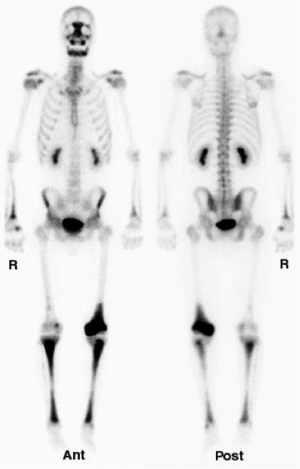
Skeletal Scintigraphy:
• bone cancer or spread of cancer to other areas can be determined
• helps to determine the location of an abnormal bone or diagnose broken bones or inflammations
Myocardial Scintigraphy:
• informs about the condition of blood flow, vitality and function of the heart muscle
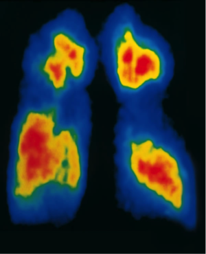
Lung scintigraphy:
• perfusion and ventilation of the lungs can be visualized
Lymphoscintigraphy:
• lymph vessels and lymph nodes are shown
Scintigraphy of Kidneys:
• renal dysfunction can be diagnosed
Scintigraphy of the thyroid gland:
• is applied for example in cases of suspected hyperthyroidism
3.Procedure
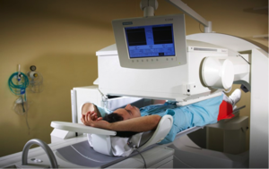
The so-called radiopharmaceutical is administered orally, injected into the bloodstream or inhaled as a gas. The radioactive labelled material accumulates in the target organ or tissue. It takes part in the metabolism without affecting or manipulating it. The radiotracer releases gamma rays. A special gamma camera is now able, under help off a special computer, to create a picture of the organ. As the camera detects the radioactive energy, the picture provides information about the function and structure of the examined organ. To indicate any possible abnormalities, it has to be said that in diseased tissue, metabolism runs faster or slower than in healthy tissue. To visualize this process, more or less of the radioactive material appears brighter or darker in the scintigram. To understand this process even better, the example of cholescintigraphy can be very useful. In this case, the radiotracer is injected intravenously into the patient. It now enters the bloodstream until the liver removes the radioactive pharmaceutical from the blood. The radiopharmaceutical takes part in the secretion into bile, which flows in the gallbladder via the bile ducts. Basically, a cholescintigraphy is carried out to diagnose an obstruction of the bile ducts by gallstones, tumors or bile leaks.
4.Conclusion
To begin with, the method of scintigraphy provides unique information about the function of tissues. Furthermore it gives very useful information that is needed for a correct diagnosis and the appropriate treatment of the patient. Concerning skeletal scintigraphy, abnormalities that could have been missed by a conventional radiograph or x-ray, are detected by this method. Therefore early treatment, which can be decisive for the cure of the patient, can be given. In addition, the early detection of primary cancer or cancer that has spread to other areas in the body is provided by scintigraphy. Concerning the disadvantages, it can be said that a small risk of cell-or tissue-damage and carcinogenesis always exists when a patient is exposed to any radiation, even if its level is very low. Allergic reactions to radiopharmaceuticals are very rare. During pregnancy, the developing foetus can be influenced by the radioactive material, which is transmitted to the baby through breast milk.
5.References - BETTER REFERENCES AVAILABLE
1.Walter J. Doll, Ph.D: What is Gamma Scintigraphy?
2.Dr. Eric Fréneaux/Dr Alain Denis Keller: Scintigraphy Scans
3.Dr. Hans Peter Sochor: Szintigraphie
4.W. Heindel: Szintigraphie
5.Dr. med. Peter Mahlknecht: Szintigraphie


