Potential between two phases
Phases of the cardiac action potential[edit | edit source]
Introduction[edit | edit source]
The cardiac action potential is by definition, a fast, local and significant change of the resting potential of the excitable cell which concerns the physiology of the electric activity of the heart.
The action potential occurs when the electrical membrane potential of a cell rises and falls in a very small amount of time. Here, we are referring to the specific case of the cardiac cell membrane. By using the term 'electric membrane potential', we are talking about the difference of electric potential between the inside and outside of a cell WHICH RESULTS FROM CHANGES IN the concentrations of ions on both sides off the cell wall. In order to study the action potential, we can divide its progress into 5 different phases.
Why is it important in clinical medicine ?[edit | edit source]
As we are speaking of cardiac cells, this topic relates to cardiology specifically. The electrical heart activity can be recorded by electrocardiography by placing electrodes on a patient's body. The depolarisation of the cardiomyocytes during each heartbeat is therefore detectable and very useful to doctors for a diagnosis in any related symptoms of heart problems of a patient. (Careful! The graph of an ECG and of the action potential of a cardiac cell are not the same).
Types of cardiac cells[edit | edit source]
In the heart, there are two types of cardiac action potentials. As Richard E. Klabunde explains on his Cardiovascular Physiology Concepts site, "[1]" these are the non-pacemaker and the pacemaker cardiac cells which are essential to the heart's special excitatory and contractile system. Both types of cells have a very similar action potential, and therefore differ to the action potentials in the CARDIAC MYOCYTES. For this article, we will study the action potentials of non-pacemaker cells of the heart such as atrial myocytes, ventricular myocytes and Purkinje cells.
The five phases[edit | edit source]
There are five phases of the cardiac NON-PACEMAKER action potential. Phases 1 to 3 are all repolarization phases and form a refractory period DURING WHICH the cell can not respond to a new stimulus.
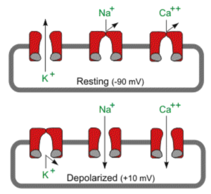
Phase 0: Rapid depolarisation. A rapid shift in membrane potential from -90mV to approximately +20mV is created by increased sodium and decreased potassium conductances.
Phase 1: Initial repolarisation. The membrane starts to repolarise slowly by decreased sodium and increased potassium conductances.
Phase 2: Plateau. The rate of repolarisation is slowed down by entering calcium, prolonging the refractory period.
Phase 3: Repolarisation. Increased conductances of potassium and a decrease of entering calcium make the potential membrane drop to approximately -90mV.
Phase 4: Resting potential. The cell is not being stimulated: the potential membrane stays between -85mV and -95mV.
Conclusion[edit | edit source]
For the heart to function without being consciously asked to by our brains, it relies on autorhythmicity. The cells need to depolarise spontaneously. Research in this area include work on the several types of cells within the heart but also all the ion channels responsible in membrane potential change. The advance in both fields help understand rhythm disorders and therefore fixing them as well, with for instance, the revolutionary invention of the pacemaker.
Links[edit | edit source]
1. Richard E. Klabunde, PhD: Cardiac action potentials
2. Greg Ikonnikov and Dominique Yelle: Physiology of cardiac conduction and contractility
3. From Emergency Medical Paramedic 2010-2013 official website: Emmergency Medical Paramedic, Cardiac Action Potential
References[edit | edit source]
- ↑ Non-pacemaker action potentials, also called "fast response" action potentials because of their rapid depolarization, are found throughout the heart except for the pacemaker cells. The pacemaker cells generate spontaneous action potentials that are also termed "slow response" action potentials because of their slower rate of depolarization. These are found in the sinoatrial and atrioventricular nodes of the heart.


