Perimetry
|
This article was marked by its author as Under construction, but the last edit is older than 30 days. If you want to edit this page, please try to contact its author first (you fill find him in the history). Watch the discussion as well. If the author will not continue in work, remove the template Last update: Friday, 16 Dec 2016 at 4.44 pm. |
|
Check of this article is requested. Suggested reviewer: Carmeljcaruana |
Introduction[edit | edit source]
What is Perimetry?[edit | edit source]
Perimetry is the systematic measurement of differential light sensitivity in a visual field. Perimetry incorporates the presence of test targets on a defined background, perimetry is just one way to systematically test the visual field (Medline, 2015)
Visual Field[edit | edit source]
The Visual field refers the overall area in which objects can be seen in the peripheral (side) vision as the eyes of the subject are focused on a central point (Medline, 2015). Furthermore visual field testing is a component of a routine comprehensive eye exam carried out by an optometrist or ophthalmologist, the test includes determining a patients full horizontal and vertical range of sensitivity of their vision (Haddrill et al, Jan 2016).
How is the Test Performed?[edit | edit source]
Visual field test are used to detect various eye diseases such as Scotomas, Optic nerve damage and Optic neuropathy (Haddrill et al, Jan 2016) among others, this paper aims to provide further information on the current literature as well as future developments and its impact on modern day medicine. Three main field and automated exams are used to perform this test, which are as follows:
Confrontation Visual Field Exam This exam is an elemental check of the visual field.
Goldmann Field Exam In the Goldman field exam the patient is located centrally 3 feet from the screen, and asked to stare at an object and report to the examiner when the object migrates into the peripheral vision.
Automated Perimetry In automated Perimetry the patient sits in front of a concave dome and stares at an object in the middle, the patient presses a button when small flashes of light are observed (Medline, 2015).
Importance in clinical medicine[edit | edit source]
Perimetry in medicine[edit | edit source]
Perimetry is a standardized, clinically established method for measuring the visual field. It is therefore of great importance in diagnostics in ophthalmology. Perimetry offers the possibility to obtain a comprehensive insight into the function and integrity of the visual system in a short time.
Role in practical medicine[edit | edit source]
In clinics, perimetry is used for the observation of ocular, retinal and neurological diseases, which are accompanied by a successive deterioration, up to the loss of the visual performance, The loss of a certain number of axons of the optic nerve lead to areas of reduced eyesight in the visual field, so called Scotomas. Modern computed Perimetry enables ophthalmologists to detect areas of reduced sensivity. Physicians can associate these defects in the visual field with certain diseases (e.g. Glaucoma), by assesing and interpreting their patterns. Perimetry, besides Tonometry, Gonioscopy and Ophthalmoscopy is of particular importance in the diagnosis and follow-up of glaucoma.Glaucoma is one of the most common causes of blindness.
Worldwide Relevance[edit | edit source]
The number of glaucoma patients is estimated to be 72,9 million in 2020, of which around 11,2 million will go blind ambilaterally, because their disease is not treated in time. Glaucoma is a slowly progressing disease, which requires the patient to be monitored regularly undergo Perimerty tests until the end of his life.
Literature review[edit | edit source]
How does it Work?[edit | edit source]
The working principle of Perimetry can be outlined using the Static Automated Perimetry procedure as a template. The perimeter is linked to a virtual program on the accompanying computer. The program tests the central 30 degrees of the visual field using a six degree spaced grid. (“What is Perimetry?” n.d) A visual sensitivity map is generated after lights of same size and color but varying intensities are flashed arbitrarily in the patient’s visual field. The patient usually indicates a response by pushing a button whenever light is seen. Thus, allowing the computer to successfully calculate the patient’s visual field.
The measurement of visual field is of great significance in Ophthalmological exams. Consequently, various Perimetry techniques have been developed. However, the two most popular types are the Automated and Manual Perimetry procedures. Automated Perimetry electronically maps a visual field while Manual Perimetry, such as the Goldmann or the Tangent Screen technique, requires trained professionals to draw out the visual field. Automated Perimetry is universally acknowledged as the standard Perimetry procedure. This is because it provides greater standardization, calibration and statistical analysis of results. (Miller, Walsh, & Hoyt, 2005). The instrument’s high sensitivity and specificity produces detailed and valid data.
Advantages and disadvantages[edit | edit source]
However, patients may find it inconvenient due to the prolonged test time and the plausible fatigue. The lack of flexibility in the procedure, such as examining only certain variables is also a major disadvantage of Automated Perimetry. To successfully avoid these setbacks, Manual Perimetry using the Goldmann meter can be carried out. Since there is a trained professional subjects can be given breaks when experiencing fatigue. Subjects would also feel encouraged to respond to the stimuli in the presence of an examiner. Certain aspects of the visual field can be specifically explored for more accurate mapping. On the other hand, this technique is less efficient, includes the possibility of human error and lacks the sensitivity of an electronic device. Furthermore, results are extremely subjective and may not be easily reproduced. Finally, examiners may not be sufficiently qualified or trained. Despite the differences in examining the visual field, both methods require the patient’s collaboration. The visual field data produced is only as reliable as the patient’s response.
The equipment[edit | edit source]
Perimeter[edit | edit source]
| Automated perimeter |
|
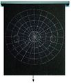
| Tangent screen |
|
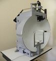
| Goldmann perimeter |
|
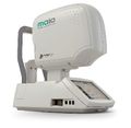
| Microperimeter |
|
The methodology[edit | edit source]
- Turn both required equipments on.
- Open the operating system.
- Create a new file for the new patient.
- After completing the patient’s personal data in the file, start a new examination.
- Determine the perimeters of the measurement.
- Choose one eye (right/left) to start the procedure.
- The non-tested eye should be covered and the patient should sit comfortably and remain concentrated in front of the perimeter screen. Silence should be kept.
- The patient must keep his eyesight fixated on the target in the middle of the screen.
- The recognition of any other lights in the screen should be done by the patient by pressing the response button.
- The examiner sets demonstration mode first, before the actual examination starts, in order for the patient to get used to the response button.
- At the end of the test, the results regarding fixation losses, false positives or negatives will be displayed on the screen.
Conclusion[edit | edit source]
Automated perimetry has evolves exponentially over recent years due in part to modern computer technology that enables more complex visual stimuli and test procedures to be realised as opposed to the traditional methods of testing (McKendrick, 2005). Advancements in Perimetry could characterize and monitor a range of optical diseases such as glaucomas, this could aid treatment and improve management of these diseases. The development and validation of these new visual field procedures can be accomplished but must adhere to multicentre trials that yield clinical significance. Perimetry and visual field testing could be enhanced as diagnostic tools in clinical practice(Johnson et al, 2015).
References[edit | edit source]
- Br J Ophthalmol., March 2016; available from: https://www.ncbi.nlm.nih.gov/pubmed/16488940
- Carroll JN, Johnson CA. Visual Field Testing: From One Medical Student to Another. EyeRounds.org. August 21, 2013; available from http://EyeRounds.org/tutorials/VF-testing/
- Foundation, G. R. (n.d.). "Five common Glaucoma Tests". Glaucoma.org. Retrieved 2014-02-20.
- McKendrick, A.M., 2005. Recent developments in perimetry: test stimuli and procedures. Clin. Exp. Optom. 88, 73–80. doi:10.1111/j.1444-0938.2005.tb06671.x
- Micro perimeter ophthalmic examination / non-mydriatic retinal camera / table - MAIA - CenterVue - Videos [WWW Document], n.d. URL http://www.medicalexpo.com/prod/centervue/product-80144-503154.html (accessed 12.11.16).
- Miller, N.R., Walsh, F.B., Hoyt, W.F., 2005. Walsh and Hoyt’s Clinical Neuro-ophthalmology. Lippincott Williams & Wilkins.
- Perimetry Test (Visual Field Testing) for Glaucoma [WWW Document], n.d. . WebMD. URL http://www.webmd.com/eye-health/perimetry-test-visual-field-testing-for-glaucoma (accessed 12.11.16).
- Quigley, Broman; „Thenumber of people with glaucoma worldwide in 2010 and 2020“;Röhmel, Diss:“Der Einfluss transkranieller Gleichstromstimulation der visuellen Kortex auf die Kontrastwahrnehmung in der Schwellenperimetrie“; Medizinische Fakultät der Charité-Universitätsmedizin Berlin, October 10, 2013
- Sides Media, www sidesmedia com, n.d. Glaucoma Today - The Next Generation in Perimetry [WWW Document]. Glaucoma Today. URL http://glaucomatoday.com//glaucomatoday/2015/12/the-next-generation-in-perimetry/ (accessed 12.11.16).
- Slonim, C., MD, n.d. Visual Field Testing [WWW Document]. Vis. URL http://www.allaboutvision.com/eye-exam/visual-field.htm (accessed 12.11.16).
- Visual field: MedlinePlus Medical Encyclopedia [WWW Document], n.d. URL https://medlineplus.gov/ency/article/003879.htm (accessed 12.11.16).
- Visual Field Perimetry Testing Equipment (Visual Field Perimeters) | OptometryWeb: The Ultimate Online Resource for Optometrists [WWW Document], n.d. URL http://www.optometryweb.com/Eye-and-Vision-Examination-Products/6411-Visual-Field-Perimetry-Testing-Equipment-Visual-Field-Perimeters/ (accessed 12.11.16).
- What is Perimetry? [WWW Document], n.d. URL http://webeye.ophth.uiowa.edu/ips/Perimetr.htm (accessed 12.11.16).


