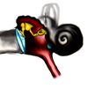
Ossicula auditus
In the middle ear cavity the smallest paired ossicles of the body can be found, namely the auditory ossicles. They create a transmission complex of vibrations from the eardrum to the fenestra vestibuli ossis temporalis, to the perilymphatic space. There are three bones:
- malleus – hammer, bone pressing on the ear drum
- incus – anvil, middle bone
- stapes – stirrup, the most medial bone adjacent to the base of the fenestra vestibuli
Anatomical structures[edit | edit source]
Malleus[edit | edit source]
Bone club-shaped, tapering caudally. We recognize several formations on it:
- caput mallei – rounded part for connection with the anvil, projecting high above the upper edge of the eardrum;
- collum mallei – neck pressing medially from the pars flaccida membranae tympani;
- manubrium mallei – handle fused with the drum in the stria mallearis;
- proc. lateralis and proc. anterior are projections, performing minor functions.
Incus[edit | edit source]
It is located between the hammer and the stirrup. It contains:
- corpus incudis – the body, which on one side is articulated with the collum mallei and on the other, tapering side with the caput stapedis;
- crus breve – a short arm extending to the wall of the aditus ad antrum, used for fixation;
- crus longum – a long shoulder in the mediocaudal direction, passes in proc. lenticularis, a cartilaginous projection connected to the head of the stirrup.
Stapes[edit | edit source]
The last of the ossicles embedded in the fenestra vestibuli:
- caput stapedis – spherical head of the stirrup attached to the cartilaginous process of the anvil;
- crus anterius et posterius – bony arms towards the base;
- basis stapedis – elongated plate fixed with lig. annulare stapedis to the fenestra vestibuli.
Connection of auditory ossicles[edit | edit source]
The auditory ossicles are connected by ligaments, sometimes with the character of a joint connection. Numerous ligaments bind them to the surroundings.
Functions[edit | edit source]
Thanks to the connection of these three bones, a transmission chain is created that perceives the vibrations of the eardrum, which are transferred to the perilymph of the labyrinth of the Inner ear. At the same time, the oscillations change from high amplitude and low intensity starting at the drum to low amplitude with high intensity. This ensures transmission to the perilymph, from there to the endolymph, to the hearing receptor itself in the cochlea.
Links[edit | edit source]
Related articles[edit | edit source]
Used literature[edit | edit source]
- ČIHÁK, Radomír. Anatomie. 2. edition. Praha : Grada, 2001. 516 pp. vol. 1. pp. 177. ISBN 978-80-7169-970-5.
- ČIHÁK, Radomír – GRIM, Miloš. Anatomie. 2.. edition. Praha : Grada Publishing, 2004. 673 pp. vol. 3. pp. 628-629. ISBN 80-247-1132-X.


