Nasal Cavity
Nasal cavity, choanae, vascular and nerve supply
Introduction[edit | edit source]
The nasal cavity serves as both an olfactory and respiratory organ and is housed within the nasal skeleton. It comprises two bony nasal cavities separated by the nasal septum, which often deviates from the midline. These cavities open anteriorly into the piriform aperture and posteriorly into the nasopharynx through structures called choanae.
The nasal cavity and septum are lined with nasal mucosa, bound to the periosteum and perichondrium of supporting bones.
Boundaries[edit | edit source]
• Roof- Nasal bone, Nasal part of frontal bone, cribriform plate and body of sphenoid
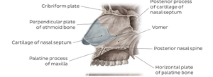
• Floor – incisive bone, palatine process of maxilla, horizontal plate of palatine bone
• Lateral wall – Frontal process of maxilla, body of maxilla, inferior nasal concha, lacrimal bone, ethmoidal labyrinth, perpendicular plate of palatine bone and medial plate of pterygoid process of sphenoid , cartilaginous nasal wall
• Medial wall – Vomer, Perpendicular plate of ethmoid, septal nasal cartilage
Relation to other structures[edit | edit source]
• Cranially – Anterior cranial fossa (via cribriform plate of ethmoid)
• Caudally – oral cavity via hard plate
• Laterocranially – orbit via ethmoidal bone
• Laterocaudally- maxillary sinus
• Ventrally – nose
• Dorsally – nasopharynx via choana
Functionally, the nasal cavity warms and humidifies inspired air, removes pathogens and particulate matter, facilitates the sense of smell, and drains paranasal sinuses and lacrimal ducts.
Nasal concha[edit | edit source]
There are 3 nasal concha: superior, middle and inferior nasal conchae
The superior and middle nasal conchae are inward bony projections originating from the ethmoid bone, extending into the nasal cavity. They contribute to the creation of three distinct spaces within the nasal cavity:
- The spheno-ethmoidal recess: it is situated above the superior nasal concha. Its function is to drain the sphenoidal sinus, which is an air-filled cavity within the sphenoid bone.
- The superior nasal meatus: Positioned between the superior and middle nasal conchae, this meatus connects with the posterior ethmoidal sinuses, which are air-filled spaces within the ethmoid bone.
- The middle nasal meatus: Formed by the superiorly located middle nasal concha and the inferior nasal concha below, the middle meatus is deeper and longer compared to the superior meatus. It serves as a passageway for drainage from various sinuses, including the frontal sinus (located in the frontal bone), via the ethmoidal infundibulum. Additionally, it facilitates drainage from the maxillary and anterior ethmoidal sinuses.
The inferior nasal concha is the largest and widest among the three nasal conchae. Unlike the superior and middle conchae, it is comprised of a separate bone known as the inferior nasal concha. Its inner surface is lined with a mucous membrane containing sizable vascular spaces, which can expand or contract to regulate the width of the nasal cavity.
Functionally, the inferior nasal concha contributes to the formation of two areas within the nasal cavity: the middle and inferior nasal meatuses.
The inferior nasal meatus, situated beneath the inferior nasal concha and the lateral nasal wall, is the largest space within the nasal cavity. It plays a crucial role in directing airflow, as well as in the processes of humidifying, heating, and filtering the air that enters through the nose. Additionally, the inferior nasal meatus receives lacrimal fluid from the nasolacrimal canal through an aperture known as Hassner’s valve.
Vascular supply[edit | edit source]
• Arterial supply – anterior and posterior ethmoidal arteries, descending palatine artery (maxillary artery), sphenopalatine artery (maxillary artery), facial artery
• Venous drainage – facial vein, ethmoidal veins, pterygoid plexus, pharyngeal plexus
Innervation[edit | edit source]
- Somatosensory – anterior ethmoidal nerve, posterior nasal branches and infraorbital nerve
- Visceromotor sympathetic – periarterial plexus and deep petrosal nerve
- Visceromotor parasympathetic – facial nerve
- Special sensory – olfactory nerve
References[edit | edit source]
- HUDÁK, Radovan – KACHLÍK, David. Memorix anatomie. 2. edition. Triton, 2013. ISBN 978-80-7387-712-5.


