M-FISH
M-FISH, or multi-color fluorescent in situ hybridization, is one of the modern applications of the basic FISH method and thus ranks among molecular cytogenetic methods.
Classic FISH[edit | edit source]
FISH works with already specific probes that hybridize with chromosomes or their parts based on the suspicion of a specific problem. Which makes it somewhat narrower and more expensive for us to use it. Therefore, in cases where we are just finding out where the problem is in the individual's genome, we use the advantages of the M-FISH method, which hybridizes all chromosomes.
Probes[edit | edit source]
Probes are stretches of nucleic acid that are variously labeled and artificially prepared. Classic FISH uses the following probes:
- Centromere, hybridizing with repetitive sequences
- Locus-specific, hybridizing to certain DNA sequences (genes)
- Painting, hybridizing with practically the entire chromosome
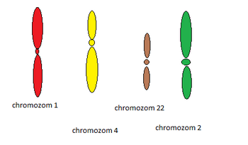
However, for the needs of M-FISH, only painting or whole-chromosome probes are used. There are 24 of them, each hybridizing with another of the chromosomes. They are marked with different combinations of fluorochromes.
Signal detection[edit | edit source]
Microscopes with filters are used for evaluation. There must be at least six of these for proper fluorochrome resolution. The signals are read by a special computer program.
Use[edit | edit source]
This method is used in molecular cytogenetics to diagnose larger previously unknown chromosome rearrangements. However, its condition still remains the cultivation of cells to observe mitotic nuclei, which slows down the speed of obtaining results.
Other similar methods[edit | edit source]
Another similar method is the SKY (Spectral karyotyping) method, which, however, uses a spectrometer to detect signals. Even the M-BAND (multicolour banding) method is based on a similar principle. Here, however, we use several differently labeled locus-specific probes. Which 1 will mark a certain chromosome with colored bars. It is suitable for identifying structural changes of a given chromosome.
Links[edit | edit source]
Used literature[edit | edit source]
- KOČÁREK, Eduard – PÁNEK, Martin. Klinická cytogenetika I : Úvod do klinické cytogenetiky. 2. edition. Karolinum, 2010. 134 pp. ISBN 978-80-246-1880-7.


