Lower limb deformities

Lower limb deformities we can divide by where they occur. They can be either congenital or acquired during life (limb overload, trauma, metabolic disease, inflammation, etc.).
Knee joint deformities[edit | edit source]
Genua valga[edit | edit source]
Genua valga (bent knees) are characterized by an increase in the physiological valgosity of the knee joints.
Genua vara[edit | edit source]
Genua vara (deviated knees) they represent a very common deformity of childhood. They are normal in newborns. When the baby starts walking, the deformity becomes even more pronounced.
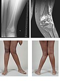
Genu recurvatum[edit | edit source]
Uncommon defect. It used to be associated with rickets, today it occurs more often during epiphyseal cartilage disorders for example by inflammation, injury or paralysis.
Clinically, hyperextension of the knee joint is present. The lower leg and thigh form an open angle forward
Small defects do not need to be treated. If the stability of the limb is disturbed, we choose a surgical solution in the form of an osteotomy in the supracondylar region of the femur, as well as surgery on the surrounding soft tissues.
Genu flectum[edit | edit source]
It occurs most often in children with cerebral palsy. In adults, it is associated with deformative osteoarthritis knee joint, which is associated with the development of a varus position of the knee, or at rheumatoid arthritis, which is associated with vaginal disability and flexion contracture.
Clinically, we see a knee in flexion, where full extension is not possible.
Therapy depends on the cause of the disease. In cerebral palsy, we choose the prolongation of tendons of the flexors of the knee joint. In adults with deformable osteoarthritis and flexion contracture, a complete joint replacement is required.
Genua valga 
Genua vara 
Genu recurvatum


Leg deformity[edit | edit source]
Pes equinus[edit | edit source]
English vertical leg. It is a deformity that occurs in disorders of the nervous system in spastic paralysis, weak polio after poliomyelitis or polio extensor.
Clinically, the foot is fixed in plantar flexion. The patient steps on the front of the foot, unable to step on the heel. Such walking leads to the collapse of the transverse arch and the formation of bruises on the tread. Atrophy of the calf and leg muscles occurs.
Treatment can be conservative, which includes rehabilitation, training of a shortened Achilles tendon, or the use of corrective plaster.
The Achilles tendon can be surgically extended.
Pes excavatus[edit | edit source]
The arched foot usually has neurological causes (myelodysplasia, muscular dystrophy), Friedreich's disease, or secondary to inflammation.
Clinically, the longitudinal arch is highlighted. There is a contracture of the plantar fascia and soft tissues in the area. The fingers tend to have a clawed position and bruises occur in the area of the metatarsal heads. The heels are varous.
Before therapy, it is necessary to send the patient for neurological examination. It can be conservatively solved with the help of special orthopedic shoes.
The surgical solution consists in releasing the plantar structures from the heel bone, or performing a wedge-shaped corrective osteotomy.
Pes planovalgus[edit | edit source]
Longitudinally flat foot. There is an abnormal reduction or disappearance of the longitudinal vault. The heel is in a valgus position, the talus lowers and the forefoot abducts.
Pes transversoplanus[edit | edit source]
Transverse flat foot usually occurs after the age of 30, more often in women, who more often choose inappropriate footwear (short tight toe). In the wrong shoes, the forefoot is overloaded, which is further exacerbated by any overweight.
Clinically, pain occurs in the landscape of metatarsal heads, sometimes neuralgic pain from n oppression. plantaris medialis (radiating between the 3rd and 4th fingers). When the load is relieved, the pain usually subsides. There is a gradual reduction of the arch by weakening of the ligaments and muscles. The front of the leg widens and the metatarsal heads arch into the area, under which bruises form.
The transverse flat foot usually occurs together with the flat foot, so therapy is usually associated with both of these deformities. Most often we choose orthopedic insoles, or hearts for shoes. You can also use physical therapy, hydrotherapy and whirlpools. For neuralgias, spraying can be used.
If the pain does not subside, it is necessary to choose a surgical solution, which consists in nerve release by crossing the anterior edge of the intermetatarsal plantar ligament. For more severe defects (rheumatic disease) we resect the headers II.-V. metatarsus.
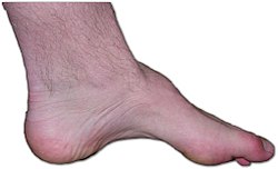
Pes excavatus 
Pes planovalgus


Toe deformities[edit | edit source]
Hallux valgus[edit | edit source]
The arched toe is one of the most common deformities in the foot. It is much more common in adults due to the weakening of the ligament and muscular system, which leads to a decrease in the transverse arch and a change in the position of the thumb.
Hallux varus[edit | edit source]
Deviated thumb. Unlike valgus, it is not so common.
Hallux rigidus[edit | edit source]
They weren't a stiff thumb. It most often occurs on the basis of osteoarthritis in the metatarsophalangeal joint of the thumb. It occurs in some professions or also after inflammation and surgery.
Digitus malleus[edit | edit source]
This deformity usually occurs in connection with hallux valgus and dog transversoplanus. We distinguish two forms, namely the hammer finger and the mallet finger.

Hallux valgus 
Hallux varus 
Hallux rigidus


Diseases in the heel area[edit | edit source]
Haglund's exostosis[edit | edit source]
Exostosis occurring on the back of the heel bone due to long-term mechanical irritation.
Calcar calcanei[edit | edit source]
Or the spur is a deformity in the heel bone. It most often occurs between the ages of 40 and 60 and is present on both sides.

Calcar calcanei 
Haglundova exostóza

.jpg)
Links[edit | edit source]
Reference[edit | edit source]
Used literature[edit | edit source]
| „ | {{{1}}} | “ |