Lens
The lens is one of the refractive structures. It is located behind the pupil in the camera oculi posterior, which is filled with fluid (humor aqueous). The lens is suspended from the inner circumference of the ciliary body (corpus cilliare). The basic property of the lens is accommodation - the ability to change optical power.
The lens of an adult:
| diameter | 9–10 mm |
| thickness | 3,7–4,4 mm |
| optical power | 10–17 dioptre |
Anatomy and histology of the lens[edit | edit source]
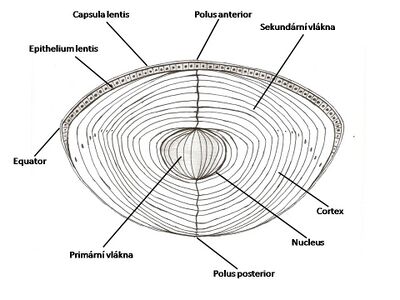
- Facies anterior lentis has a spherical curvature.
- Facies posterior lentis has a parabolic curvature.
- Equator lentis forms the circumference of the lens.
- Polus anterior is the most ventral point of the lens.
- Polus posterior is the most distal point of the lens.
- Axis lentis is the axis of the lens.
- Capsula lentis or lens capsule is the transparent membrane that encases the entire lens, but it is thicker on the anterior side than on the posterior side and is thickest at all at the periphery of the lens. It is produced by the anterior epithelium and consists of amorphous glycoproteins and collagen IV.
- Epithelium lentis or anterior subcapsular epithelium of the lens is a single-layered cuboidal epithelium. The individual cells are interconnected by nexuses and form numerous interdigitations with the lens fibers. The cells change into fibres towards the periphery of the lens.
- Fibrae lentis are the fibres of the lens and are shaped like a hexagonal prism. The fibers reach lengths of up to 7-10 mm. They contain proteins called crystallins. The fibres are arranged in such a way that there are visible seams at the front and back. It is Y-shaped on the front and inverted Y-shaped on the back. The fibres clamp into one seam, continue towards the equator, flip to the opposite side and clamp to the other seam.
- Nucleus lentis is the deep and stiff component of the lens.
- Cortex lentis is made up of the more superficial fibres, which are arranged in such a way that sutures are visible on the anterior and posterior sides. It is shaped like a Y on the front and an inverted Y on the back. The fibres are clamped in one seam, continue towards the equator, fold over to the opposite side and clamp to the other seam. The closer to the center the fibers start on one side, the farther from the center they attach on the other side, implying that the fibers in one layer are the same length.
Lens development - embryology[edit | edit source]
On the 22nd day of development, the optic sulcus on the sides of the future forebrain appears for the first time. They are growing and turning into an optic vesicles. These vesicles come into contact with the surface epithelium (ectoderm) and induce the formation of the lens plate. From this plate, the lens originates. The plate gradually slips in and forms the lens pit. During the 5th week, the pit loses contact with the superficial ectoderm and the lens vesicle is formed. Shortly after its formation, the cells on the posterior wall of the vesicle begin to stretch and change into long fibres. As the fibers increase in number and gradually stretch, they soon begin to fill the lumen of the lens vesicle, completely filling it by the end of the 7th week. The cells at the front of the vesicle form the anterior cuboidal epithelium of the lens.
Links[edit | edit source]
Related articles[edit | edit source]
Literature[edit | edit source]
- ČIHÁK, Radomír. Anatomie 3. 2. edition. Grada, 2004. 692 pp. vol. 3. ISBN 978-80-247-1132-4.
- KONRÁDOVÁ, Václava – UHLÍK, Jiří – VAJNER, Luděk. Funkční histologie. 2. edition. H & H, 2000. 291 pp. ISBN 80-86022-80-3.
- SADLER, Thomas, W – SINHA, M.D. Langmanova lékařská embryologie. 1. české edition. Grada, 2011. 414 pp. ISBN 978-80-247-2640-3.

