Lateral neck region, scalenic fissure

Under construction / Forgotten
|
This article was marked by its author as Under construction, but the last edit is older than 30 days. If you want to edit this page, please try to contact its author first (you fill find him in the history). Watch the discussion as well. If the author will not continue in work, remove the template Last update: Thursday, 06 Feb 2025 at 2.53 pm. |
Lateral neck region has the following borders:
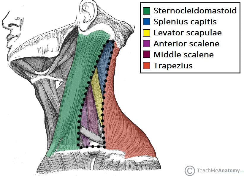
- Posterior - trapezius muscle
- Inferior - clavicle bone
- Roof - investing layer of deep cervical fascia
- Floor - prevertebral fascia, scalene muscles, levator scapulae muscle, splenius capitis muscle

The inferior belly of omohyoid muscle subdivides it into 2 triangles: “Omoclavicular triangle” & “Omotrapezius triangle”.
Omoclavicular triangle:
Also called “supraclavicular/ subclavian triangle”
Borders:
- Anterior - sternocleidomastoid muscle
- Superior - inferior belly of omohyoid muscle
- Inferior - clavicle bone
- Floor - anterior and medial scalene muscles, 1’st rib, 1’st dictation of serrated anterior
- Roof - skin, fascia, platysma muscle
Contents:
- Subclavian artery, suprascapular, transverse cervical, which are branches of the thyrocervical trunk
- Subclavian vein
- Phrenic nerve
- Brachial plexus
- External jugular vein
- Fibres of cervical plexus
- Supraclavicular lymph node

Omotrapezius triangle:
Also called “Occipital triangle”
Borders:
- Anterior- sternocleidomastoid muscle
- Posterior- trapezius muscle
- Inferior- inferior belly of omohyoid muscle
- Roof- skin, superficial & deep cervical fascias
- Floor- splenius capitis muscle, levator scapulae, middle and posterior scalene muscles
Contents:
- Erb’s point- “Punctum Nervosum”. Lesser occipital nerve, great auricular nerve, transverse cervical nerve, supraclavicular nerve
- Accessory nerve
- Phrenic nerve
- External jugular vein
- Transverse cervical artery
- Transverse cervical vein

Scalene fissure:
The gap between anterior and middle scalene muscles. Located deep in the lateral cervical region
Borders:
- Anterior- anterior scalene muscle
- Posterior- middle scalene muscle
- Inferior- 1’st rib
Contents:
- Brachial plexus
- Subclavian artery
Scalenovertebral triangle:
Borders:
- Medial- longus colli muscle, vertebral column, esophagus, thyroid gland and trachea
- Lateral- anterior scalene muscle
- Superior- “carotid tubercle” (anterior tubercle of C6 on the transverse process which might compress the common carotid artery)
- Inferior- 1st rib
- Roof- skin and platysma
Contents:
- Venous angle- point of drainage of both thoracic duct and right lymphatic duct. Between the internal jugular and subclavian vein
- Stellate ganglion- the connection of 1st thoracic and inferior cervical ganglion
- Middle cervical ganglion
- Thyrocervical trunk- transverse, superficial cervical artery and ascending cervical artery
- Vertebral artery
- Thoracic duct
Sources
Stingl, J., Grim, M., & Druga, R. (2012). ''Regional anatomy''. Galen.

