Hydrocephalus (neonatology)


Hydrocephalus is an excessive accumulation of cerebrospinal fluid with enlargement of the ventricular system of the brain. Hydrocephalus occurs as a result of an imbalance between the production and absorption of cerebrospinal fluid or as a result of an obstacle in its natural circulation. It can be accompanied by increased intraventricular pressure.[1]
'Ventriculomegaly is an enlargement of the brain's ventricles. The cause may be increased intraventricular pressure (as in hydrocephalus) or passive enlargement of the ventricles during brain atrophy. Causes of fetal ventriculomegaly include: abnormal cerebrospinal fluid circulation, agenesis of the corpus callosum, neuronal migration disorders (lissencephaly, schizencephaly), neuron proliferation disorders (megalencephaly, microcephaly), holoprosencephaly, abnormalities of cerebral vessels. Fetal ventriculomegaly is often associated with syndromes based on chromosomal defects.[2]
Pathophysiology[edit | edit source]
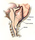
Cerebrospinal fluid flows from the lateral ventricles through the foramen of Monro (foramen interventriculare) into III. chambers, from there through the aqueductus Sylvii (aqueductus mesencephali) to IV. chambers and further through the foramina of Luschkae (aperturae laterales ventriculi quarti) and Magendie into the subarachnoid space, where it is absorbed into the venous circulation via the arachnoid villi that line the superior sagittal sinus.
Under normal circumstances, CSF is formed at a rate of 0.3-0.4 ml/min. (500 ml/day). The total volume of cerebrospinal fluid is 40 ml in full-term and 10-30 ml in premature newborns. The mean opening pressure of the cerebrospinal fluid is 10 cmH2O in term infants and 9.5 cmH2O in premature infants.[2]
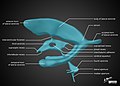
Anatomy of the ventricular system.

Circulation of cerebrospinal fluid.


Classification[edit | edit source]
- non-communicating hydrocephalus – obstruction of the circulation of the cerebrospinal fluid (historically: the contrast agent applied to the ventricular system of the brain did not pass into the lumbar subarachnoid spaces;
- communicating hydrocephalus – obstruction of external cerebrospinal fluid pathways, hypersecretion or hyporesorption of cerebrospinal fluid.[1]
Etiology[edit | edit source]
- 'congenital hydrocephalus
- stenosis of the Sylvian ureter – the most common congenital cause; arises spontaneously or during TORCH infections, CNS malformations or as part of some genetically linked syndromes;
- Arnold-Chiari malformation – congenital structural defect of the cerebellum with expansion of the brain tissue, possibly and the brainstem to the foramen magnum and the upper level of the cervical spine;
- Dandy-Walker syndrome – aplasia of the vermis of the cerebellum and a large cyst in the posterior cranial fossa, widely open to IV. chambers, atresia of holes IV. chambers;
- neural tube defects – meningomyelocele;
- arachnoid cyst;
- arteriovenous malformation of the vein of Galen;
- hydrancephaly – damage to the end brain in the basin of the internal carotid artery; the cerebral hemispheres consist only of a thin membrane filled with cerebrospinal fluid;
- postinfectious hydrocephalus – bacterial meningitis and arachnoiditis or ventriculitis;
- posthemorrhagic ventriculomegaly and hydrocephalus.[1]

Arnold-Chiari malformation type I – herniation of the cerebellar tonsils into the foramen magnum (MRI of the brain).
Chiari malformation II. type.

Variant of Dandy-Walker syndrome - pontine and cerebellar dysplasia (MRI, T2-weighting).

Spinal fissures.


Clinical picture[edit | edit source]
- normotensive hydrocephalus – asymptomatic;
- tension hydrocephalus – signs of intracranial hypertension:
- increasing head circumference (> 2 cm/week, > 2 cm above 97th percentile for gestational age), tense large fontanelle, cranial suture spacing;
- apnoeic pauses, bradycardia, hypertension, respiratory disorders;
- gastrointestinal symptoms: reduced sucking reflex, food intolerance, vomiting;
- symptom of the "setting sun" - deviation of the bulbs caudally; nystagmus, convergent strabismus (paresis of the VI cranial nerve);
- sudden change in behavior (apathy/irritability), impaired consciousness;
- convulsions, rigidity;
- Cushing's triad: bradycardia, hypertension, wide pulse pressure.[1]
Diagnosis[edit | edit source]
- prenatal (ultrasound);
- clinical picture;
- ultrasound (ventricular index according to Levene);
- cerebrospinal fluid examination;
- MRI, CT.[1]
Treatment[edit | edit source]
- temporary: lumbar puncture (withdrawal of 10 – 15 ml/kg), subcutaneous reservoir, external ventricular drainage (ventriculostomy);
- surgical – shunt operation: ventriculoperitoneal shunt; endoscopic surgery (for non-communicating hydrocephalus): ventriculostomy III. chambers, obstruction of the Sylvian ureter, septostomy.[1]