Glandula thyroidea (SFLT)
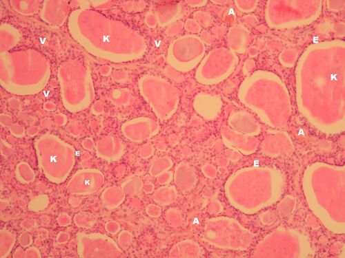
Glandula thyroidea - (HE)[edit | edit source]
Description: K - colloid (thyreoglobulin), E - monolayer epithelium of follicles, V - connective stroma, A - capillary.
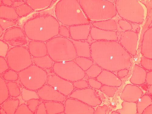
Glandula thyroidea - (HE)[edit | edit source]
Description: F - follicle filled with colloid, E - monolayer epithelium of follicles, V - connective stroma.
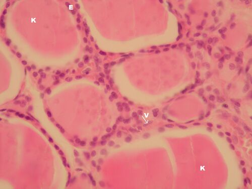
Glandula thyroidea - detail (HE)[edit | edit source]
Description: K - colloid, E - monolayer epithelium of thyroid follicles, V - connective tissue forming fine bands around follicles, arrow points to fibroblast nucleus.
Note: The colloid precipitates into the middle of the follicles when the sample is processed into a slide and then breaks off during slicing. These are artifacts.
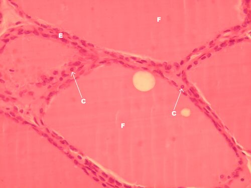
Glandula thyroidea – detail (HE)[edit | edit source]
Description: F - follicle formed by monolayer epithelium (E) and filled with colloid, C - probably parafollicular cells (C cells - calcitonin producers).
Glandula thyroidea – detail (HE)[edit | edit source]
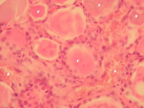
Description: F - follicles formed by monolayer epithelium, the height of which correlates with thyroid activity (from flat in hypofunction to cylindrical in hyperfunction), here cubic epithelium (= eufunction), A - capillaries in the connective tissue stroma (capillaries have fenestrated endothelium).


