Fluorescence microscope
A fluorescence microscope is an optical microscope that uses fluorescence to image both organic and inorganic structures. The first fluorescence microscope was constructed around 1911 by Carl Reichert.
In fluorescence microscopy, shorter wavelengths in the UV radiation area are used, when the substance absorbs ultraviolet rays and emits visible light of longer wavelengths, which can then be observed with a light microscope. The atom is irradiated, where under normal circumstances electrons orbit the nucleus in layers (orbitals) with a certain energy level. During UV radiation, electrons absorb energy and are excited (excited) to a higher energy level. Electrons are unstable at a higher energy level and return back to the ground state. The movement of an electron from a higher energy level to a lower one is accompanied by the emission of a photon.
Structure and function of a microscope[edit | edit source]
The source of radiation is usually a high-pressure mercury vapor lamp. The quartz tube contains a small amount of mercury and an inert gas. In the interval 356–546 nm, it emits the greatest amount of energy. The power of the discharge lamps varies, discharge lamps with a power of 50, 100 and 200 W are used. The excitation radiation first passes through an excitation filter. This filter only allows light of the aforementioned wavelength to pass through. It prevents the penetration of light with the wavelengths of radiation emitted (fluorescence), which would cause an undesirable colored background. Next, the light falls on a dichroic mirror, set at an angle of 45°.
Epifluorescence microscope[edit | edit source]
The radiation is reflected and passes through the objective to the examined sample. It causes the excitation of electrons in the sample and the subsequent emission of light. The emitted radiation has a longer wavelength and spreads in all directions. Thanks to the immersion objective and the oil, only a part of it passes through and also hits the dichroic mirror. This mirror removes unwanted excitation radiation of high intensity (dangerous to the eye, evaluation of observation) and transmits the emitted radiation. The overall efficiency of separation of excitation and emitted radiation by the dichroic mirror is more than 80%. The emitted radiation then passes through a barrier filter. This only allows the emitted radiation in the length according to the fluorochrome used, shorter wavelengths stop it. Furthermore, we can observe fluorescent radiation with an eyepiece or capture it with a camera.
Transmitted-light fluorescence microscope[edit | edit source]
It is also used in laboratories, but has a different layout. The source of radiation is placed under the specimen in the same way as in a classic microscope. A specially arranged shading condenser then allows the light to fall on the sample from the side. The dangerous excitation light then flies outside the lens and only the emitted light passes through the lens. The epifluorescence type of microscope is currently more popular than the transmission type.
Primary fluorescence (autofluorescence, natural fluorescence)[edit | edit source]
This type of fluorescence occurs only in some cells (mainly in plant cells, tissues) and the fluorescence intensity is not very strong. In the area of UV radiation, it is primarily bound to proteins/amino acids – tryptophan, phenylalanine, tyrosine, and in the area of visible light to reduced forms of NADH, NADPH, vitamin A, chlorophyll, cytochromes, hemoglobin, myoglobin.
Secondary fluorescence[edit | edit source]
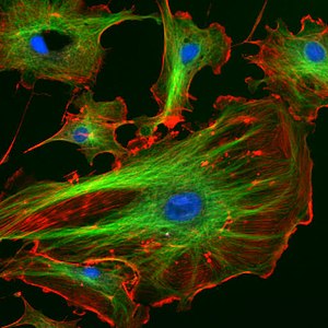
This type consists in applying a fluorescent substance (fluorochrome) to the examined structures, which allows us to visualize them after irradiation. Known fluorochromes for the visualization of nucleic acids include, for example, acridine orange, ethidium bromide or DAPI (4', 6-diamidino-2-phenylindole dihydrochloride). Also FITC (fluorescein-5-isothiocyanate), TRIC, Texas Red.
Among the widely used markers is the green fluorescent protein GFP (green fluorescent protein), which was isolated from the seaweed Aequrea victoria and thanks to which we can monitor marked structures in the cell. In addition to green fluorescent protein, its yellow and blue-green variants and the unrelated red fluorescent protein are also available today.
Use[edit | edit source]
Fluorescence microscopy was the first to allow us to visualize the structures of the cytoskeleton of eukaryotic cells, it is used in immunohistology, immunocytology and immunocytochemistry. In cell biology, it is widely used to identify components of the cytoskeleton, cell organelles or to monitor biochemical events. In microbiology, a fluorescence microscope is used to identify different bacterial genera. In cytogenetics, the FISH (in situ hybridization) method is used, when using a fluorescently labeled DNA (RNA) probe, we detect certain selected sections of DNA or chromosomes and can diagnose, for example, chromosomal aberrations, microdeletions, microscopic breaks, etc.
Gallery[edit | edit source]
Links[edit | edit source]
References[edit | edit source]
- ↑KOČÁREK, Eduard a Martin PÁNEK. Klinická cytogenetika I : úvod do klinické cytogenetiky. 2. vydání. Praha : Karolinum, 2010. 134 s. ISBN 978-80-246-1880-7.
- ↑ HAMPL, Vladimír, et al. Fluorescenční mikroskopie [online]. Poslední revize 4.10.2012, [cit. 2013-02-05]. <http://web.natur.cuni.cz/~parazit/parpages/mikroskopickatechnika/fluorescencni.htm>.
Sources[edit | edit source]
- VYMĚTALOVÁ, Veronika. Biologie pro biomedicínské inženýrství. I. díl. 1. vydání. Praha : Česká technika - nakladatelství ČVUT, 2008. ISBN 978-80-01-04013-3.


