File:Rectus sheath.jpg
From WikiLectures
Rectus_sheath.jpg (263 × 297 pixels, file size: 27 KB, MIME type: image/jpeg)
Summary[edit | edit source]
- Description
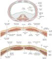
- Relationship between rectus sheath and the parietal peritoneum and transversalis fascia. Section through the abdominal wall superior to the arcuate line. Section through the abdominal wall inferior to the arcuate line.
- Author
- Schuenke M, Schulte E, Schumacher U. THIEME Atlas of Anatomy. General Anatomy and Musculoskeletal System. Illustrations by Voll M and Wesker K. 3rd ed. New York: Thieme Medical Publishers; 2020.
- Source
- : 3.2.2 Rectus Sheath. In: Wikenheiser J, Voll M, Wesker K, ed. Clinical Anatomy, Histology, Embryology, and Neuroanatomy: An Integrated Textbook. 1st Edition. New York: Thieme; 2022.
- Date
- 27.03.2024
File history
Click on a date/time to view the file as it appeared at that time.
| Date/Time | Thumbnail | Dimensions | User | Comment | |
|---|---|---|---|---|---|
| current | 20:01, 27 March 2024 |  | 263 × 297 (27 KB) | Shaked.Fru (talk | contribs) | {{File |description = Relationship between rectus sheath and the parietal peritoneum and transversalis fascia. Section through the abdominal wall superior to the arcuate line. Section through the abdominal wall inferior to the arcuate line. |author = Schuenke M, Schulte E, Schumacher U. THIEME Atlas of Anatomy. General Anatomy and Musculoskeletal System. Illustrations by Voll M and Wesker K. 3rd ed. New York: Thieme Medical Publishers; 2020. |source = : 3.2.2 Rectus Sheath. In: Wike... |
You cannot overwrite this file.
File usage
The following page uses this file:
