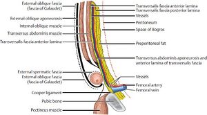
Abdominal wall - Function, Muscles, fascias
Abdominal wall - Function, Muscles, fascias[edit | edit source]
The abdominal wall encloses the abdominal cavity.
It can be divided into the anterolateral section and the posterior section.
Function of the abdominal wall[edit | edit source]
The abdominal wall functions to:
- protect the internal abdominal organs (abdominal viscera)
- Stabilization and rotation of the trunk
- Increase of intra-abdominal pressure (forceful expiration, coughing, vomiting).
Anterolateral abdominal wall[edit | edit source]
The Anterolateral abdominal wall consists of four main layers:
- Skin
- Superficial fascia
- Muscles (and associated fascia)
- Parietal peritoneum
Muscles of the Anterolateral abdominal wall[edit | edit source]
The muscles in the Anterolateral abdominal wall can be divided into 2 groups:
- Flat muscles-
- The fibers of the flat muscles run in different directions and cross each other, decreasing the likelihood of the abdominal viscera herniating through them.
- In the medial aspect (anteromedial area), each flat muscle creates an aponeurosis (a broad, flat tendon), covering the rectus muscle. The aponeurosis of all of the flat muscles is the Linea alba (a fibrous structure extending from the xiphoid process of the sternum to the pubic symphysis).
- Vertical muscles
Flat muscles:[edit | edit source]
| Name | Origin | Insertion | Innervation | Function |
| External oblique
(Fibers run inferomedially) |
Ribs 5-12 | Iliac crest and pubic tubercle | Thoraco-abdominal nerves (T7-T11) and subcostal nerve | Contralateral rotation of torso |
| Internal oblique
(Fibers run superomedially) |
Inguinal ligament, iliac crest, lumbodorsal fascia | Ribs 10-12 | Thoraco-abdominal nerves (T7-T11) and subcostal nerve | Bilateral contraction compresses the abdomen
Unilateral contraction causes ipsilateral rotation of torso |
| Transversus abdominis
(Fibres run transversely) |
Inguinal ligament, costal cartilages 7-12, iliac crest, thoracolumbar fascia | Xiphoid process, pubic crest and linea alba | Thoraco-abdominal nerves (T7-T11) and subcostal nerve | Compression of abdomen |
Vertical muscles:[edit | edit source]
| Name | Origin | Insertion | Innervation | Function |
| Rectus abdominis | Pubic crest | Xiphoid process and costal cartilages 5-7 | Thoraco-abdominal nerves (T7-T11) | Compresses abdominal viscera and stabilises pelvis during walking |
| Pyramidalis | Pubic crest and pubic symphysis | Linea alba | Subcostal nerve | Tenses the linea alba |
Mnemonic to remember the anterolateral abdominal wall muscles:
TIRE Pump
Transversus abdominis, Internal oblique, Rectus abdominis, External oblique, Pyramidalis
Fascias of the Anterolateral abdominal wall[edit | edit source]
The skin is the most superficial layer of the anterolateral abdominal wall.
The superficial fascia is located just below the skin, and consists of connective tissue.
Superficial fascia[edit | edit source]
The superficial fascia can be seen both superiorly and anteriorly to the umbilicus, and has different composition in each part.
Above the umbilicus- The superficial fascia is continuous and is made of one layer.
Below the umbilicus- The superficial fascia divides into 2 layers:
- Superficial Camper’s fascia- A thick fatty layer. Its thickness varies.
- Deep Scarpa’s fascia- A thinner and more dense membranous layer. It overlyies the muscle layer of the abdominal wall and attaches to the Linea alba and Pubic symphysis. It fuses with the Fascia lata (deep fascia of the thigh) below the inguinal ligament.
Posterior abdominal wall[edit | edit source]
Muscles of the posterior abdominal wall[edit | edit source]
The posterior abdominal wall is supported by the 12th thoracic vertebrae as well as all 5 lumbar vertebrae.
There are between 3 to 4 muscles in the posterior abdominal wall, depending on the individual: Quadratus lumborum, Psoas major, Iliacus, Psoas minor (Psoas minor muscle is only present in 40% of the population).
Only the quadratus lumborum is a ‘true’ posterior abdominal muscle, as all other muscles extend into the lower limb.
| Name | Origin | Insertion | Innervation | Function |
| Quadratus lumborum | Iliac crest and iliolumbar ligament | Transverse process of L1-L4 and inferior border of rib 12 | Anterior rami of T12-L4 | Extension and lateral flexion of vertebral column. |
| Psoas major | Transverse processes and vertebral bodies of T12-L4 | Lesser trochanter of femur | Anterior rami of L1-L3 | Flexion of thigh.
Lateral flexion of vertebral column. |
| Iliacus | Iliac fossa | Lesser trochanter of femur | Femoral nerve L2-L4 | Thigh flexion and external rotation.
Trunk flexion. |
| Psoas minor | Originates from the vertebral bodies of T12 and L1 | Pectineal line of pubis | Anterior rami of L1 | Flexion of vertebral column. |
Fascias of the posterior abdominal wall[edit | edit source]
The fascias of the posterior abdominal wall lie immediately below the skin and subcutaneous tissue.
Thoracolumbar fascia[edit | edit source]
A large, diamond shaped area of connective tissue. It is formed by the thoracic and lumbar parts of the deep fascia.
It is divided into 3 layers: Anterior, Middle, posterior.
- The Quadratus femoris muscle is enclosed between the anterior and middle layers.
- The deep back muscles are enclosed between the middle and posterior layers.
Psoas fascia[edit | edit source]
The psoas fascia is more profound to the anterior layer of the thoracolumbar fascia, and it covers the Psoas major muscle.
It is continuous with the thoracolumbar fascia laterally and with the iliac fascia inferiorly.
Sources
[1] Abdominal wall. (n.d.). Kenhub.
[2] Jones, O. (2012). The Anterolateral Abdominal Wall - Muscles - TeachMeAnatomy. Teachmeanatomy.info.
[3] Muscles. (n.d.). Google Docs. Retrieved March 27, 2024 - Salvador, Rishi and Anurag's notes


