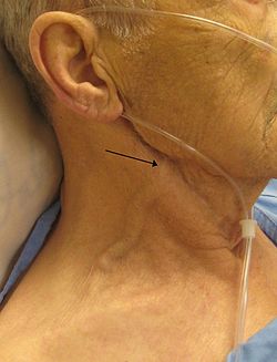
Examination of the neck
Neck examination consists of inspection, percussion and auscultation. The shape and length of the neck, motility, jugular veins, arteries, lymph nodes and thyroid gland are examined.
Shape and length[edit | edit source]
The shape and length should correspond to the physical proportions of the individual. In obese people, the neck is relatively larger. On the other hand, the neck is usually thin in malnourished people with a "sunken" appearance.
Mobility[edit | edit source]
We ask the patient to lean forward and tilt his head and look to the sides. Physiologically, the neck should be mobile in all directions.
Anteflexion[edit | edit source]
We ask the patient to tilt his head forward. Normally, the individual should be able to touch his chest with his chin. However, especially in older people, they might often be unable to do so due to degenerative spinal disease. If the patient is unable to have neck anteflexion, it may be a sign of neck opposition, which is usually caused by meningitis or subarachnoid hemorrhage.
Artery[edit | edit source]
Inspection[edit | edit source]
- Visible pulsation of the external carotid artery - during exertion, hypertension and thyrotoxicosis
Palpation[edit | edit source]
- We feel the pulsation of the external carotid artery that runs beneath the sternocleidomastoid muscle
- If we find a decreased or even disappearance of the external carotid pulsation unilaterally (only on one side), it probably indicates a narrowing of the carotid artery by the thrombus
- If we find a decrease or even disappearance of the external carotid pulsation bilaterally (on both sides), it may be a narrowing of the common carotid artery or aortic valve
Ascultation[edit | edit source]
- We ask the patient to inhale, exhale and hold his breath for a while (so that we do not hear the air flow through the trachea)
- We focus on murmurs that are audible during an aneurysm or narrowing of the carotid artery, or are transmitted from the aortic orifice (in aortic stenosis)
Veins[edit | edit source]
Inspection[edit | edit source]
- We observe the filling of the jugular veins (venae jugulares externae), which roughly indicates the central venous pressure
- We examine the patient's exhalation when he is lying on his back with his head resting, which he turns to the side. The filling of the jugular veins should not be increased by more than 2 cm above the reference plane, which is a horizontal plane intersected by the sternoclavicular joint
- When pressing on the right hypochondrium, if we observe an increased filling of the jugular veins (due to the hepatojugular reflux), it could suggest hepatopathies or right heart failure
- Collar of Stokes - compression of the superior vena cava leads to the expansion of venous plexuses with edematous infiltration of the neck, possibly also of the head and upper limbs
Lymph nodes[edit | edit source]
Palpation[edit | edit source]
Lymph nodes are not palpable under physiological circumstances.
We examine the following lymph nodes:
- occipital,
- suboccipital,
- preauricular,
- retroauricular,
- behind the edge of the mandible,
- submandibular,
- submental,
- cervical,
- supraclavicular.
When palpable, we evaluate the size, consistency, pain and motility of the lymph nodes. Lymph node enlargement can be caused by inflammation or tumors. Inflammatory nodules are softer, painful and mobile against the surroundings. On the other hand, the nodules affected by the tumor are stiffer, painless, immobile against the surroundings and could form aggregrations.
Thyroid gland[edit | edit source]
The thyroid gland is not palpable or visible under physiological conditions. It is only palpable and visible when when there is an enlargement (= goiter)
We evaluate its:
- size
- consistency - soft, stiff, hard stones
- mobility against the skin
- symmetry
- sensitivity to touch
An enlarged thyroid gland can oppress the esophagus and cause swallowing difficulties. It could also compress the trachea and cause breathing difficulties. Moreover, it can put pressure on the recurrent laryngeal nerve, leading to dysphonia (disorders of the voice).
Palpation[edit | edit source]
We feel the thyroid gland with the head tilted (due to the relaxation of the neck muscles) in the lower 1/3 of the sternocleidomastoid muscle. We feel either from behind with both hands or from the front. We also notice the movement when the patient swallows.
Goiter[edit | edit source]
It can be diffuse or nodular. Diffuse goiter is usually bilateral, but may involve only one lobe or may affect only the isthmus. Nodular goiter could involve one nodule (uninodular) or more than on nodule (multinodular).


