Electroencephalography
Electroencephalography (EEG) is a diagnostic method used to record the electrical activity of the brain. EEG is one of the non-invasive methods. Changes in the polarisation of neurons are sensed by surface electrodes.
EEG principle[edit | edit source]
Electroencephalogram is a record of the time change of polarisation of neurons and neuroglia in the CNS. It is mainly the activity of surface structures (the effect of subcortical ones on the record is much smaller), the amplitude of potentials from the surface of the skull skin in tens of μV (membrane potential in mV). The source of EEG activity is mainly excitatory (EPSP) and inhibitory postsynaptic potentials (IPSP), and significantly less AP (although they are larger, but much shorter and not so often). Pacemaker-type neurons are particularly important for EEG genesis - spontaneous production of oscillating discharges, inhibitory interneurons and feedback connections → basic principle of oscillator function (cortical neural networks → rhythmic activity 10-40Hz).
The basis of synchronised EEG activity are AP volleys → mass EPSP on cortical neurons; the essence of synchronous. Discharges of thalamic nuclei: change of their membrane potential (← reciprocal innervation with nucleus reticularis thalami, their GABA-ergic inhibitory interneurons hyperpolarize the membrane of thalamic transfer neurons) → incoming information (EPSP) → activation of voltage Ca2+ channels → change of membrane potential by input of other calcium ions → Ca2+ channels → trigger level → activity of thalamic neurons - AP series → membrane hyperpolarization restored by calcium-induced potassium flux → cycle repetition. The membrane potential of thalamic transfer nuclei close to the threshold is maintained by cholinergic input from the brainstem and forebrain, the same input reduces activity of nucleus reticularis thalami → prevents the induction of hyperpolarization → allows the transfer of sensory information to the cerebral cortex in the waking state.
Technically, the EEG recording compares the potential of two points on the skin of the skull = bipolar recording, or the difference in electrical potential between the active point of the brain tissue (under the active, exploratory electrode) against the point with zero potential (under the inactive, reference electrode - eg auricle, root nose) = unipolar record.
Method[edit | edit source]
The electrodes are placed evenly on the surface of the skull according to the prescribed schemes (eg. system 10-20). The electrodes are marked with letters (A = Ear lobe; C = Central; P = Parietal; F = Frontal; O = Occipital; T = Temporal) and numbers (odd numbers for electrodes located above the left cerebral hemisphere, even numbers for electrodes above the right hemisphere ). The number of scanning electrodes corresponds to the number of recording channels and the scanning method. Unipolar and bipolar connections are used. In the case of a bipolar connection, the potential difference between the two active electrodes is sensed; in the case of a unipolar connection, the sensed voltage is detected between the active electrode and the reference electrode, or clamp. For unipolar, a distinction is made between directional connections, front-rear and left-right connections. When connected, a combination of directions may also occur. Surface or subsurface electrodes can be used. Surface electrodes are used for non-invasive sensing of electrical activity of the brain from the surface of the head. Either individual electrodes or electrode caps are used. Subsurface electrodes are used for invasive sensing in electrocorticography. They can be in the form of wires, needles or targets made of a suitable material (Pt, Ag-Cl, etc.). Cotton wicks in salt solution can also be used. The conductive environment in the case of subsurface electrodes is body fluids, in the case of surface electrodes conductive gels are usually used.
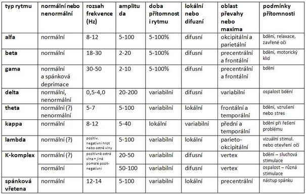
The electroencephalograph amplifies the signals and filters out the noise. It records the obtained results in a graph. Brain activity varies with the frequency and amplitude of the waves. The basic types of activities (EEG rhythms) include:
Evoked potentials[edit | edit source]
Evoked potentials are significant changes in the EEG signal caused by some external stimulus (light, sound or somatosensory). Simultaneously with the stimulus, a mark must be created in the EEG record that defines the time the stimulus occurred for later evaluation of the record.
These are synchronized responses of groups of neurons to afferent excitations or direct el. irritation = a more complex type of response than the unit activity of individual neurons. For individual potentials, we evaluate the shape, latency of peaks, amplitude, slope, polarity and mutual relations of waves. They consist of:
- primary components - electrical response of a group of first activated neurons
- late components - follows the primary, it is a response to impulses from primary neurons, or a response to afferentiation by slower fibres, has greater latency, slower course, lower amplitude, more diverse shape
Evoked potentials allow us to map projections from the periphery in trunk, subcortical and cortical structures and assess the degree of functional development of sensory systems after birth. Evoked brainstem potentials can be extracted from the EEG - this can be used to examine this area. It is much easier to record cortical evoked potentials (most pronounced in the primary projection areas of large sensory systems).
Examples:
- VEP = Visual Evoked Potential
- AEP = Auditory Evoked Potential
- BERA = Brainstem Electrical Responsy Audiometry
- CERA = Cortical Electrical Responsy Audiometry
- SSEP = Somato-Sensory Evoked Potential
Uses[edit | edit source]
Most often in neurology and psychiatry. Monitoring and diagnosis of diseases: epilepsy, coma, migraines, CNS in children. Sensed signals of electrical activity of the brain can also be used to control various devices and equipment (so-called neurofeedback) - for example, in affected patients or in the military.
Electrocorticography[edit | edit source]
Sensing the signal directly from the cerebral cortex is called electrocorticography, it is used in neurosurgery. Electrocorticography is more accurate than electroencephalography because EEG attenuates the signal as it passes through the skull, on the order of microvolts.
Links[edit | edit source]
Related articles[edit | edit source]
External links[edit | edit source]
References[edit | edit source]
- HRAZDIRA, Ivo a Vojtěch MORNSTEIN. Lékařská biofyzika a přístrojová technika. 1. vydání. Brno : Neptun, 2001. ISBN 80-902896-1-4.
- MYSLIVEČEK, Jaromír. Základy neurověd. 2. vydání. Praha : Triton, 2009. ISBN 978-80-7387-088-1.


