Elastic artery (histological slide)
From WikiLectures
Histological description[edit | edit source]
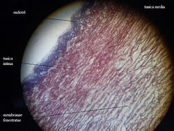
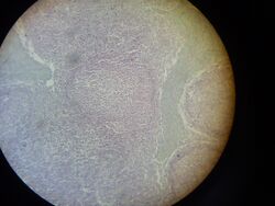
The wall of an elastic artery consists of three basic layers: Tunica intima , T. media , T. adventitia , examples of elastic arteries are large arteries (Aorta, A.subclavia, Arteria carotis communis and others.).
Tunica intima
Endothelium (single-layer squamous epithelium), lamina elastica interna.
Tunica media
In this layer, parallel arranged elastic membranes (light in HE staining) and layers of smooth muscle (dark) alternate, smooth muscle cells have a nucleus in the center (visible only at maximum magnification), lamina elastica externala is difficult to distinguish.
Tunica adventitia
It contains numerous vasa vasorum .
Links[edit | edit source]
Related articles[edit | edit source]
- Muscle artery (histological preparation, WRF)
- Muscular artery (histological slide, HE)
- Vein (histological slide, HE)
Bibliography[edit | edit source]
- MUDR. EIS, Václav – MUDR. JELÍNEK, Štěpán – MUDR. ŠPAČEK, Martin. Histopatologický atlas [online]. ©2006. [cit. 15.04.2010]. <http://histologie.lf3.cuni.cz/histologie/atlas/index.htm>.
- JUNQUIERA, L. Carlos – CARNEIRO, José – KELLEY, Robert O.. Základy histologie. 1. edition. Jinočany : H & H 1997, 1997. 502 pp. ISBN 80-85787-37-7.


