Developmental disorders of the ear

The outer and middle ear develop separately from the inner ear. Therefore, we can divide congenital ear defects into congenital defects of the outer and middle ear and congenital defects of the inner ear.
Congenital defects of the external and middle ear[edit | edit source]
Abnomalies of the external and middle ear are related to the maldevelopment of the first gill slit and the first two gill arches . These diseases are manifested by shape deformities of the auricle and ear canal. They can occur together with dysostoses of the lower jaw, zygomatic bone and auditory ossicles .
Congenital malformations of the auricles relate to shape, size and position.
Otapostasis[edit | edit source]
The most common congenital defect is otapostasis , the so-called standing bolt. It is an inherited, frequent defect of the auricle, the essence of which is the smoothing of the fold of the antihelix. The defect can be corrected surgically (the most suitable time for correction is between 5 and 6 years). The principle is the creation of a new antihelix.
Microcia[edit | edit source]
Another very common defect is microcia, i.e. reduction of the auricle. The defect can be classified into 3 degrees, in the case of the 2nd and 3rd degrees, the bolt is not only reduced in size, but also deformed in shape.
Anocia[edit | edit source]
Anocia is the complete failure of the auricle to develop.
Appendices preauriculares[edit | edit source]
Appendices preauriculares are usually unilateral appendages of the pinna . They mostly have the shape of bumps or pedunculated lobes located in front of the tragus.
Congenital ear fistula[edit | edit source]
These are canals mainly in the ascending part of the helix, external fistulas are less common, on the neck they are colo-auricular fistulas. Their danger lies in possible secondary infection, so they need to be surgically removed.
Stenosis and atresia of the ear canal[edit | edit source]
Stenosis is a narrowing of the ear canal. Atresia is a complete closure of the ear canal - the ear canal is completely impassable. Atresia can be both bony and fibrous. It is the cause of conductive hearing loss. Both defects (stenosis and atresia) can be associated with other defects (auricle disorder, middle ear disorder, facial nerve disorder ). The therapy is surgical . It consists in the canalization of the ear canal, or the reconstruction of the middle ear.

Failure to form an antihelix

Antihelix after reconstruction
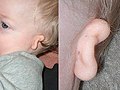
Detail of MICROTIA (outer ear deformity).

This newborn with an atretic right ear, is displaying an anomaly included in VACTERL Association. V


Congenital defects of the inner ear[edit | edit source]
- Anomalies of the inner ear are caused by disorders of otocyst formation. Different types of labyrinth hypoplasia arise due to a disorder in the development of the otocyst.
The Mondini Anomaly[edit | edit source]
- A normal snail has 2.5 turns. In the case of Mondini anomaly , the cochlea has only 1 turn and there is a pathological communication between the scala vestibuli and the scala tympani with simultaneous expansion of the saccus and ductus endolymphaticus .
- Hearing impairment varies in extent.
- Diagnosis is mainly based on CT .
Complex congenital disabilities[edit | edit source]

The ear is often affected within specific syndromes. The best known is the so-called Treacher-Collins syndrome .
Treacher-Collins syndrome[edit | edit source]
It is an AD disease. Mental development is not affected in this syndrome. Typically found here:
- antimongoloid position of eye slits with coloboma,
- hypoplasia of cheekbones and mandible ("bird profile"),
- malformation of the ear,
- macrostomia,
- high floor.
Links[edit | edit source]
Related articles[edit | edit source]
Zdroj[edit | edit source]
- BENEŠ, Jiří. Studijní materiály [online]. ©2007. [cit. 2009]. <http://jirben2.chytrak.cz/materialy/orl_jb.doc>.
References[edit | edit source]
- KLOZAR, Jan, et al. Speciální otorinolaryngologie. 1. edition. Praha : Galén, 2005. pp. 224. ISBN 80-7262-346-X.