Comparison of microscopic techniques
(Redirected from Comparison of microscopic techniques/physical principle)
Comparison of microscopic techniques[edit | edit source]
The technical field of using microscopes is called microscopy. All microscopes are used to magnify objects that cannot be seen by the naked eye. In the field of Microscopy there are three big branches of microscopic techniques. These are the optical (INCLUDING FLUORESCENCE), electron (TRANSMISSION AND SCANNING) and ATOMIC FORCE microscopes.
Optical microscopes [edit | edit source]
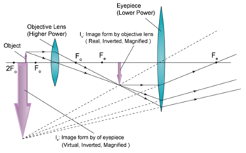
The traditional microscopes that have been used since around the 17th century are known as optical or light microscopes. The light microscope uses a set of lenses to magnify images of given samples. All optic microscopes share the same basic componets in which the light passes from the light source to the user's eyes. The lightsource or sometimes a mirror is used. The diaphragm and condenser. The objective lenses and the objective turret. The eyepiece or ocular lens. All of the magnification of light happens between the objective lens and the eyepiece. By placing the object slightly BEYOND the focal point of the first lens, the first convex lens forms an image inside the focal LENGTH of the eyepiece. the image of the first lens becomes a VIRTUAL object for the second lens, and therefore forming a very large virtual inverted image.
Fluorescence microscopy[edit | edit source]
A Fluorescence microscope is an optical based microscope used to study organic or inorganic substances. Fluorescence microscopes are based on the epifluorescence microscopy.
Fluorescence is the emission of light by a substance that has absorbed light or other radiation. The light source emittes light that passes though the objective lens and is focused on the sample.
The principle of fluorescence microscopy works as follows. The sample is illuminated by a source with a specific wavelength that can be absorbed by fluorphores. Fluorophore is a chemical compund that can re-emit light after it has absorbed light initially. Then a spectral emission filiter is used to separate the illuminated light source from the re-emited light of the flurescence. Examples for such light sources are mercury vapor lamps or high power LEDs.
Electron Microscopy[edit | edit source]
A beam of electrons if used to create a surface image of the scanned sample. When working with an electron microscope an beam of electron is used to create an surface image of a sample. The are two types of electron microscopes, the transmission electron microscope (TEM) and the scanning electron microscope (SEM).
The TEM shoots a high voltage beam of electrons through the sample to obtain information about the structure. Whereas the SEM obtains an image by a process called raster scanning. Here the specimen of a sample scanned with electrons that when interact with the surface of the given sample, producing various signals that can be detected. A digital image is then produced of the surface area of the specimen with a magnignification of up-to 500 000 times.
Atomic force microscopy[edit | edit source]
Atomic force microscopy or (AFM) is a high resolution type of scanning.
Unlike other forms of microscopy the AFM makes use of van-der-Vaals forces to produce a surface image. THOUGH THE LATERAL RESOLUTION OF AFM IS LOW (SAY 30nm) THE VERTICAL RESOLUTION CAN BE 0.1nm. The atomic force microscopy (ATM) measures the surface of a sample through a noscopically small needle that is attached to a cantilever. A cantilever is plate like structure, anchored at only one end to a support from which it is protruding.
The needle is guided line by line a defined griD across the surface of the sample. This procedure is called scanning. The bending and deflection of the tip during scanning the surface is measured and creates an image.
References[edit | edit source]
- ↑ Abramowitz M, Davidson MW (2007). "Introduction to Microscopy". Molecular Expressions. Retrieved 2007-08-22.
- ↑ Voie, A.H. (1993). "Imaging the intact guinea pig tympanic bulla by orthogonal-plane fluorescence optical sectioning microscopy"
- ↑ Duarte FJ (1993), Electro-optical interferometric microdensitometer system, US Patent 5255069.


