Cervical Spondylosis
Cervical spondylosis is a degenerative disease of the cervical spine . It is probably the result of degenerative changes in the intervertebral discs due to aging. Changes in the articular surfaces, hypertrophy of the ligamentum flavum and ossification of the posterior longitudinal ligament often occur. This can lead to the oppression of sensitive structures such as nerves and spinal cord . The disease can present with neck and shoulder pain , headache and headache , radicular symptoms and spondylogenic cervical myelopathy (SCM) . However, not all patients with radiological cervical spondylosis have clinical difficulties.
Epidemiology[edit | edit source]
Cervical spondylosis is the most common cause of spinal cord injury in patients over 55 years of age. Based on radiological findings, degenerative changes in the cervical spine were found in 90% of men over 50 and 90% of women over 60. The progression of cervical spondylosis can be very slow and the patient may be asymptomatic for a long time. Occurrence is equally common in both sexes, however, it occurs earlier in men.
Pathophysiology[edit | edit source]
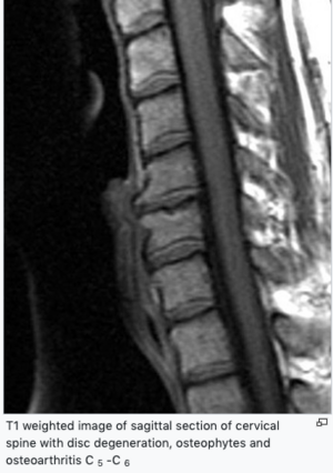
Cervical spondylosis is the result of degeneration of the intervertebral disc . As the intervertebral disc ages, it loses water, collapses and fragments. The process begins in the nucleus pulposus of the intervertebral disc. While the central annular disc attaches centrally, the annulus fibrosus on the circumference arches outward. This results in an increased mechanical load on the cartilaginous surface of the joint along the edge of the vertebral body. The vertebral body responds by subperiosteal bone formation and osteophyte formationwhich can oppress nerve tissue in the ventral part of the spinal canal. The predisposition for the development of spondylogenic cervical myelopathy is a congenital narrowing of the spinal canal (10–13 mm). Similarly, osteophytes can propagate to intervertebral foramines and irritate nerve roots. Osteophytes are probably the result of the body's efforts to stabilize the hypermobile intervertebral joint due to disc degeneration. T1 weighted image of sagittal section of cervical spine with disc degeneration, osteophytes and osteoarthritis C 5 -C 6 Ossification of the posterior longitudinal ligament may occur in the Asian population , which may contribute to the oppression of nerve structures.
Ligamentum hypertrophy of the flavum occurs with increasing age . Kyphosis and subluxation occur quite frequently , which may contribute to the SCM picture . The SCM image depends on many factors. These are static mechanical oppression, dynamic mechanical oppression, spinal cord ischemia and injuries due to tensile and shear forces. By static factors are meant, for example, a simple narrowing of the ducts due to osteophytes. Dynamic factors are directly related to static factors. Due to the movement of the spinal cord in the narrowed spinal canal, nervous tissue is irritated. During spinal anteflexion, the spinal cord is irritated by osteophytes of the ventral part of the canal. In retroflexion of the spinehypertrophic ligamentum flavum arches into the spinal canal, which compresses the spinal cord against osteophytes formed ventrally. Ischemia probably plays a role in the SCM picture. Histopathologically, in the cases of SCM, changes in the gray matter were demonstrated, while the white matter remained. This occurs in ischemia, which in this case is probably due to a microcirculatory disorder. Due to narrowing of the spinal canal and abnormal movements of nerve structures in the spinal canal in patients with SCM, nerve structures can be injured by tension and shear forces and cause local axonal damage.
Clinical picture[edit | edit source]
There are several syndromes associated with cervical spondylosis:[edit | edit source]
Cervicalgia (Intermittent neck and shoulder pain )[edit | edit source]
Cervicalgia is very common in clinical practice . However, it can be a big problem for doctors to diagnose the cause, especially if there are no other neurological symptoms in the picture. If the patient has a neurological finding, we can use imaging methods to diagnose the cause. On the other hand, having a radiologically positive finding and no neurological symptoms is usually of little informative benefit for frequent abnormalities of the cervical spine even in asymptomatic patients. The problem is also that it is not known what is the source of the pain. The syndrome is probably caused by compression of the sinovertebral nerves and the medial branch of the rami posterioris in the cervical area.
Cervicalgia is often accompanied by neck stiffness propagating to the shoulders and head, which can be chronic or episodic with long periods of remission.
One-third of patients with cervicalgia have cervical spondylosis, headache has more than two-thirds, unilateral or bilateral shoulder pain , and a significant proportion of these patients have pain in the arm, forearm, or hand .
Chronic suboccipital pain[edit | edit source]
The headache associated with cervical spondylosis is not fully understood . The head is sensitively innervated by the roots C2 – C3 , so we could expect that the pain is associated with degenerative changes in the relevant joints. However, headache in cervical myelopathy is not associated with joint degeneration .
Radiculopathy[edit | edit source]
As a result of cervical spondylosis, radiculopathy occurs most often on nerve roots C6 and C7 . It is clinically manifested by pain , paresthesias and weakness in the neck , shoulder , upper limb and interscapular space . Atypically, pain may manifest as pain in the neck (pseudoangina) or chest pain. Cervical radiculopathy is usually not associated with myelopathy.
Spondylogenic cervical myelopathy _[edit | edit source]
Spondylogenic cervical myelopathy is the most common cause of non-traumatic paraparesis or quadruparesis.[edit | edit source]
First, cervical myelopathy manifests itself in stiff neck or stabbing pain in the hand. Compressive cervical myelopathy of C3 – C5 is manifested on the hands by stiff and clumsy fingers . Patients complain of difficult writing and loss of skill, as well as weakness and sensitivity disorders in general (there may be a loss of proprioception and vibrational sensation or asymmetry in sensation of pain and heat) . A motor deficit in the upper limbs often accompanies a similar deficit in the lower limbs. Compressive myelopathy of the lower segments of the cervical spine is typically manifested by weakness , and stiffnessloss of proprioception in the lower limbs , which may show signs of spasticity . Babinsky's reflex tends to be positive . The lower limbs are usually asymmetrical .
Lhermitte's symptom may be present when the head is tilted .
Bladder congestion , urgency or incontinence may rarely occur in connection with cervical myelopathy .
More than a third of patients with cervical myelopathy suffer from anxiety or depression . This is related to limited momentum.
Central Spinal Syndrome[edit | edit source]
This syndrome typically occurs in the elderly after spinal hyperextension with a narrowed spinal canal. During the extension of the spine, the spinal cord is compressed against the ventral osteophytes, and in addition, it pushes the hypertrophic ligament flavum dorsally. Central spinal cord syndrome is accompanied by muscle weakness higher in the upper limbs than in the lower limbs and sensory disturbances below the lesion site . It can manifest itself similarly to myelopathy with spasticity and urinary retention.
Diagnostics[edit | edit source]
It is based on anamnesis and clinical findings .
Imaging methods play an important role in the diagnosis of cervical spondylosis (see below).
Determination of cyanocobalamin ( vitamin B12 ) and rapid reagin response can help distinguish the metabolic and infectious cause of myelopathy from spondylogenic.
EMG can also be diagnostically beneficial , especially for the exclusion of peripheral neuropathies. EMG is suitable for the diagnosis of radiculopathy. We can read from the EMG how long the lesion lasts.
Display methods[edit | edit source]
MRI is the method of choice for cervical spondylosis . MRI is a non-invasive non-radiation method that displays the spinal cord and subarachnoid spaces very well. Although the method is excellent for nerve structures, some spondylotic changes may escape attention (eg smaller lateral osteophytes).
A simple X-ray is an inexpensive method of detecting spondylogenic disorders in symptomatic patients. A simple image shows well the narrowing of the intervertebral disc space, osteophytes, the width of the spinal canal, etc.
CT is preferably used for better imaging of bone structures . It is good, for example, for imaging intervertebral foramines. It is sometimes used to supplement MRI scans.
Myelography or CT-myelography may be preferred .
Differential diagnostics[edit | edit source]
Amyotrophic lateral sclerosis[edit | edit source]
- Ankylosing spondylarthritis
- Cluster headache
- Migraine
- Multiple sclerosis
- Syringomyelia
- Tension headache
- Torticollis
Therapy[edit | edit source]
Treatment of cervical spondylosis consists of neck immobilization (soft collar, Philadelphia collar and others), pharmacological treatment (NSAIDs, TCAs, muscle relaxants, opioids for moderate to severe pain), lifestyle modifications (avoidance, relaxation techniques) and physiotherapy ( exercises, traction and various manipulations, which can loosen various adhesions in the spinal canal, reduce compression, improve circulation, etc.).
In some cases, a surgical solution is used . Surgical care consists of anatomical correction of degenerate structures that oppress nerve roots or spinal cord ( decompression ). The indication for surgery is unbearable pain , progressive neurological deficit or proven oppression of the nerve root or spinal cord , which are the cause of the progression of symptoms. Surgical methods will not resolve cervicalgia or suboccipital pain. In spondylogenic cervical myelopathy, the surgical solution is controversial, but it can help.
Links[edit | edit source]
Related Articles[edit | edit source]
- Myelopathy
External links[edit | edit source]
Treatment of spondylogenic cervical myelopathy (http://www.neurologiepropraxi.cz)[edit | edit source]
- Pictures for cervical spondylosis
- Degenerative diseases of the spine
References[edit | edit source]
- ↑ RANA, Sandeep S. Diagnosis and Management of Cervical Spondylosis [online]. © 2011. [feeling. 2012-01-23]. < https://emedicine.medscape.com/article/1144952-overview >.
- ↑ AL-SHATOURY, Hassan Ahmad Hassan. Cervical Spondylosis [online]. © 2012. [feeling. 2012-02-02]. < https://emedicine.medscape.com/article/306036-overview >.
- ↑Jump up to:a b c RANA, Sandeep S. Diagnosis and Management of Cervical Spondylosis [online]. © 2011. [feeling. 2012-01-23]. < https://emedicine.medscape.com/article/1144952-overview >.
- ↑Jump up to:a b c d e f g h i j k l m RANA, Sandeep S. Diagnosis and Management of Cervical Spondylosis [online]. © 2011. [feeling. 2012-01-23]. < https://emedicine.medscape.com/article/1144952-clinical >.
- ↑ ČIHÁK, Radomír and Miloš GRIM. Anatomy February 3, edited and supplemented. Prague: Grada, 2004. 673 pp. 3. ISBN 80-247-1132-X .
- ↑Jump up to:a b c RANA, Sandeep S. Diagnosis and Management of Cervical Spondylosis [online]. © 2011. [feeling. 2012-01-23]. < https://emedicine.medscape.com/article/1144952-clinical#a0217 >.
- ↑Jump up to:a b c d e RANA, Sandeep S. Diagnosis and Management of Cervical Spondylosis [online]. © 2011. [feeling. 2012-01-23]. < https://emedicine.medscape.com/article/1144952-workup >.
- ↑ RANA, Sandeep S. Diagnosis and Management of Cervical Spondylosis [online]. © 2011. [feeling. 2012-01-23]. < https://emedicine.medscape.com/article/1144952-workup >.
- ↑ RANA, Sandeep S. Diagnosis and Management of Cervical Spondylosis [online]. © 2011. [feeling. 2012-01-23]. < https://emedicine.medscape.com/article/1144952-workup >.
- ↑ RANA, Sandeep S. Diagnosis and Management of Cervical Spondylosis [online]. © 2011. [feeling. 2012-01-23]. < https://emedicine.medscape.com/article/1144952-differential >.
- ↑ RANA, Sandeep S. Diagnosis and Management of Cervical Spondylosis [online]. © 2011. [feeling. 2012-01-23]. < https://emedicine.medscape.com/article/1144952-treatment >.
- ↑ RANA, Sandeep S. Diagnosis and Management of Cervical Spondylosis [online]. © 2011. [feeling. 2012-01-23]. < https://emedicine.medscape.com/article/1144952-treatment >.

