Cell membrane - structure and function
Cell Membrane – Structure and Function
All eukaryotic cells are enveloped by a limiting membrane composed of:
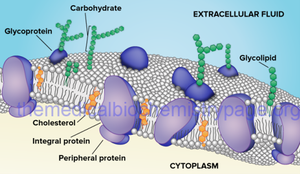
• Lipids - Phospholipids and Cholesterol, glycolipids
• Proteins
• Chains of oligosaccharides (small number of simple sugars, monosaccharides) linked to phospholipid and protein molecules.
Characteristic of biological membrane :
• Asymmetric
• Fluid Mosaic
Membranes exhibit a thickness ranging from 7.5 to 10 nm, rendering them visible solely under an Electron Microscope. They possess a trilaminar organization consisting of two hydrophilic heads surrounding a layer of hydrophobic tails.
• Phospholipids within the membrane feature two non-polar hydrophobic tails attached to a charged polar hydrophilic head group, imparting stability to the membrane due to their amphiphilic nature.
• Cholesterol is also present in membranes in a 1:1 ratio with phospholipids. It inserts itself among the fatty acid tails of phospholipids, restricting their movement.
• Additionally, membranes contain proteins known as integrins, which connect to both cytoplasmic cytoskeletal filaments and extracellular matrix components. Through these linkages, there is a continuous exchange of influences in both directions between the extracellular matrix and the cytoplasm.
Proteins of membrane, synthesized by RER, can be divided into:
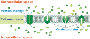
• Integral transmembrane proteins – transfer of chemical substances by channels, transporters or pumps.
1. Transporters(Carriers) \ Channels – diffusion by gap junctions of small ions, molecules, water
2. Pumps – active transport of specific ions across the membrane using energy e.g. Na+\K+ pump
3. Transport using vesicles – endocytosis and exocytosis
4. ABC Transporters – using ATP for molecules transfer, connection to MDR:
MDR (Multi drug resistance). Chemotherapy resistance emerges when cancers previously responsive to treatment begin to proliferate. In essence, the cancer cells become resilient to the effects of chemotherapy. It's not uncommon to hear remarks such as "chemotherapy for cancer was ineffective." In such cases, altering the drugs becomes necessary, as the protein responsible for transporting the drug across the cell wall ceases to function, leading to resistance of the cancer cells to the medication.
• Peripheral proteins display a less tightly bound association with one of the two surfaces of the membrane, whether inner or outer. This results in a distinct distribution of membrane proteins across the two surfaces, exhibiting an asymmetric characteristic.
The outer surface of the cell is adorned with a carbohydrate-rich region known as the glycocalyx. This layer is composed of carbohydrate chains linked to the membrane's proteins and lipids. The glycocalyx plays a crucial role in cell recognition and attachment to other cells and extracellular molecules.
• The membrane possesses a fluid mosaic appearance, attributable to the fluid nature of the lipid bilayer combined with the mosaic arrangement of membrane proteins.
Cell membrane functions:
1. The selective barrier regulates the flow of materials into and out of the cell, maintaining a constant ion concentration and thereby regulating the intracellular environment.
2. It performs a variety of specific recognition and regulatory functions.
3. These include interactions between the cell and its environment, such as adhesion and cell-to-cell signaling.
Endocytosis: Materials can traverse the plasma membrane through a general process termed endocytosis, which involves the folding and fusion of the membrane to form vesicles encapsulating the transported material.
• Phagocytosis, also known as "cell eating," involves engulfing and removing matter such as bacteria, protozoa, and dead cells. White blood cells like macrophages and neutrophils surround the bacterium with their membrane, fuse with it, and enclose it within a phagosome.
• Pinocytosis, or "cell drinking," allows small particles to enter the cell by forming an invagination, which then becomes suspended within vesicles that are subsequently broken down by lysosomes.
• Receptor-mediated endocytosis, also referred to as "absorptive pinocytosis," enables cells to absorb metabolites, hormones, and proteins by inward budding of plasma membrane vesicles containing proteins with receptor sites.
In Exocytosis, a vesicle merges with the plasma membrane, leading to the discharge of its contents into the extracellular space without compromising the integrity of the plasma membrane, which stands in contrast to endocytosis.


