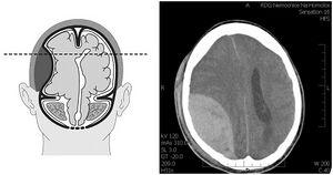
Acute Epidural Hematoma/PGS/Diagnostics
It usually results from a fracture of the calvarium at the site of the meningeal media , which is associated with rupture of the vessel. The bleeding is fast and forms an expanding mass between the dura mater and the calva (see picture). Increasing intracranial pressure results in the displacement of central structures ( uncal , tentorial or occipital herniation ) with brainstem oppression . The so-called lucid interval is sometimes anamnestic in the clinical picture, when post-traumatic loss of consciousness (brain commotion) is followed by a progressive disorder of consciousness (hematoma expansion, strain involvement) in a few minutes to tens of minutes. In addition to a characteristic anamnesis , the diagnosis is led by the finding of topical symptoms of herniation (eg Griesinger's symptom , unilateral areactive mydriasis from oppression of the oculomotor nerve in the incisura tentoria during temporal herniation) and the finding of lenticular hypertension under the calvarium on the CT head.

