File:Tuberculosis-x-ray.jpg
From WikiLectures
No higher resolution available.
Tuberculosis-x-ray.jpg (700 × 542 pixels, file size: 32 KB, MIME type: image/jpeg)
Summary
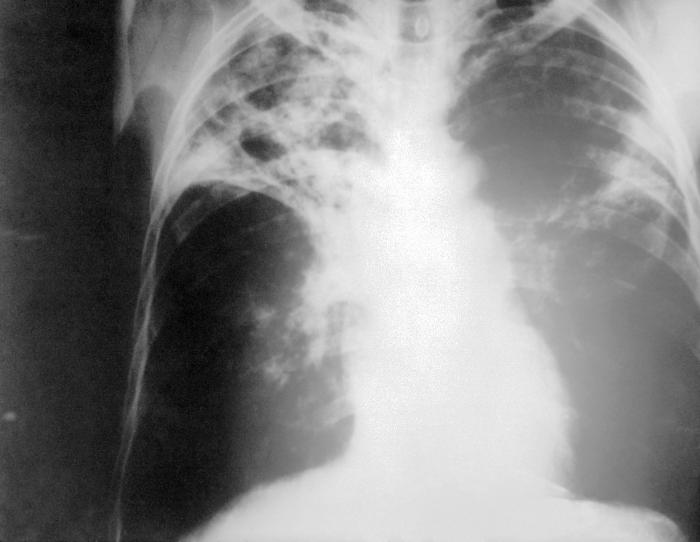
| Description |
English: An anteroposterior X-ray of a patient diagnosed with advanced bilateral pulmonary tuberculosis. This AP X-ray of the chest reveals the presence of bilateral pulmonary infiltrate, and „caving formation“ present in the right apical region. The diagnosis is far-advanced tuberculosis.
Deutsch: Eine Röntgenaufnahme im anterior-posterioren Strahlengang eines Patienten, bei dem eine beidseitige Lungentuberkulose festgestellt wurde. Diese Thorax-Aufnahme zeigt beidseitige Lungeninfiltrate und eine sogenannte „Kaverne“ im rechten Oberfeld. Sie entspricht der Diagnose einer fortgeschrittenen Lungentuberkulose. |
||
| Date |
|
||
| Source |
|
||
| Author |
|
||
| Permission (Reusing this file) |
PD-USGov-HHS-CDC English: None - This image is in the public domain and thus free of any copyright restrictions. As a matter of courtesy we request that the content provider be credited and notified in any public or private usage of this image. |
Licensing
This image is a work of the Centers for Disease Control and Prevention, part of the United States Department of Health and Human Services, taken or made as part of an employee's official duties. As a work of the U.S. federal government, the image is in the public domain.
|
Original upload log
The original description page was here. All following user names refer to en.wikipedia.
- 2005-06-05 14:21 Rsabbatini 700×542×8 (32649 bytes) An anteroposterior [[X-ray]] of a patient diagnosed with advanced bilateral [[lung|pulmonary]] [[tuberculosis]]. This AP X-ray of the [[chest]] reveals the presence of bilateral pulmonary infiltrate, and “caving formation” present in the right apical
File history
Click on a date/time to view the file as it appeared at that time.
| Date/Time | Thumbnail | Dimensions | User | Comment | |
|---|---|---|---|---|---|
| current | 14:19, 20 June 2006 | | 700 × 542 (32 KB) | wikimediacommons>Der Lange | {{Information |Description= *'''en''': An anteroposterior X-ray of a patient diagnosed with advanced bilateral pulmonary tuberculosis. This AP X-ray of the chest reveals the presence of bilateral pulmonary infiltrate, and „caving formation“ present in |
File usage
The following page uses this file: