Construction and function of a light microscope


A light microscope is a complex optical device that, with the help of several optical systems, enlarges the eye of vision and thereby improving its Resolution up to a thousand times.
- Components of a light microscope and their functions
A modern light microscope consists of three basic optical systems:
These parts are supplemented by a mechanical system into one functional unit. In developmentally older microscopes, it was common to use only one biconvex connecting lens and a kahan or candle light [1].
The light microscope is mainly used for its relatively easy production and the ability to observe preparations dynamically, without damaging them (unlike microscopes using other types of electromagnetic radiation) and with preservation of color (unlike electron microscopes).
In the preparation of specimens, dyes are often used , which cover the true color of the sample; however, different shades and depths of color are still preserved in places with different chemical and physical properties.
Lighting apparatus[edit | edit source]
The lighting apparatus is used to illuminate the object plane as perfectly as possible (that is, the plane of the table on which the specimen is located). The main function is to ensure the correct direction and intensity of the light rays to adequately illuminate the preparation (without unwanted reflections, so that the features and structures of the preparation are as clear as possible). This function is best fulfilled when applying the so-called Köhler lighting principle, which will be described below.
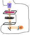
Diagram of light flow through a light microscope: 1) light source, 2) collector lens, 3) mirror, 4) field diaphragm, 5) condenser and condenser diaphragm, 6) stage with specimen, 7) objective
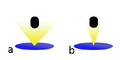
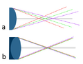
Diagram of the appearance of the cone of light when the condenser diaphragm is open (a) and closed (b) - the condenser is located at the bottom, the light is directed into the lens.
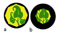
Schematic of the appearance of almost completely open (a) and half-closed (b) field diaphragms when viewed through the eyepiece.


The lighting apparatus usually consists of:
- a light source, usually a halogen lamp with a power of 50-100 W or a xenon lamp with a power of 75-150 W,
- collector lens, which concentrates the light rays coming from the source,
- field diaphragm, which is located just behind the collector lens at the light source and determines the diameter of the column of light going into the condenser; with a half-closed field diaphragm we will see a part of the image with half the diameter, with a fully open field diaphragm we will see almost the entire image [2],
- with a condenser and condenser (aperture) screen - the condenser concentrates the light in a symmetrical cone towards the specimen; the condenser aperture adjusts the angle of the rays at the top of the cone: when the condenser aperture is fully open, the angle of the rays is larger, the microscope image loses color depth and contrast, it is brighter and with better resolution; when the condenser diaphragm is closed, the beam angle is smaller and the image acquires color depth and contrast, but at the same time darkens and becomes indistinct [3] - the best results are achieved if only the field of view of the lens is illuminated.
After being directed by the lighting apparatus, the light first passes through the observed specimen and then through the objective and eyepiece. Angled mirrors can be placed between the individual components of the lighting apparatus, which direct the light rays in the desired direction, but do not adjust the shape of the light cylinder/cone.
Köhler's Illumination Principle[edit | edit source]
Köhler's lighting principle is the principle of setting the light apparatus in such a way as to achieve the best possible results in the contrast of the specimen. Applying Köhler's principle, the condenser projects a field diaphragm into the object plane, and the condenser diaphragm allows light to flow only into the field of view of the objective [4].
Lens optical system[edit | edit source]
The objective is the most important part of the microscope - the quality of the objective determines the resulting magnification of the microscope and the resulting image quality. It is also the most difficult to construct. It usually contains a large number of lenses of different shapes and in different groups (in triplets, doublets or individually) fixed in the lens barrel. The layout, number and shape of the lenses is individual for each type of lens and significantly affects all lens parameters. The entire lens system of the objective functions together as a connecting lens.
In total, we distinguish three basic construction types of lenses: achromatic lens, fluorite lens and the most complex apochromatic lens. Achromatic and fluorite lenses have a smaller number of lenses at the same magnification value and thus a significantly worse correction of optical defects [5].

Basic lens parameters [6][edit | edit source]
These parameters fundamentally depend on the construction of the lens. In practice, the values for a specific lens are usually written on the side of the body (mount) of the lens itself.
- The numerical aperture of the lens, which depends on the index of refraction of the lens and the aperture angle (the angle of the rays at the apex of the cone of light emerging from the condenser diaphragm, see Lighting apparatus) by the following relationship:
- NA = n * sin (α),
- where NA is the Numerical Aperture, n is the Refractive Index and α is the Aperture Angle; the numerical aperture is directly proportional to the microscope's distinguishing feature, and is therefore one of the most fundamental characteristics of the objectives - we can easily increase it by using a front plano-convex lens in the construction of the microscope (which increases the aperture angle) or by adding immersion oil (which increases the refractive index [7];
- focal length of the lens, determined by the distance of the object focal point of the lens from the object principal plane; the focal length affects the magnification of the objective (and thus the overall magnification of the microscope);
- magnification of the objective (see Zvětšení mikroskopu), which is determined by the focal length of the objective, the objective provides transverse magnification (another component of the overall magnification of the microscope is the angular magnification of the microscope, see Eyepiece optical system);
- lens aperture, which describes how much the lens resists passing light; is determined by the ratio of the focal length f to the diameter of the cone of light emerging from the condenser diaphragm (see Lighting apparatus); ideální světelnost objektivu je f:1, the ideal lens aperture is f:1 , practically the best lens aperture is around f:1.4 [8];
- correction of optical defects.
| Value | Mark | Unit | Essential for: |
|---|---|---|---|
| numerical aperture | NA | - | resolution limit of a microscope (direct proportionality) |
| focal length of the lens | f | m [meter] | transverse lens magnification |
| lens magnification | Z | - | overall magnification of the microscope |
| lens aperture | k | - | finding the optimal power of light to create a clear image |
Optical lens defects [9][edit | edit source]
- Spherical aberration occurs when light rays passing through the periphery of the lens and through the center of the lens cross at different points, creating multiple foci; this is one of the most harmful disorders that strongly affects the resolution and quality of the resulting image; for the correction of spherical aberration, apertures (which reduce the number of rays falling on the periphery of the lens) and front and meniscoid objective lenses, which have a minimal peripheral area of the lens, are used;
- chromatic aberration occurs as a result of the different refractive index of glass for different wavelengths (individual colors of the spectrum) − depending on its wavelength, each colored ray emerges at a different angle and intersects with a ray of the same color at a different point, thus similar to spherical aberration the formation of several foci; the result of chromatic aberration is a point of light "circled" by the colors of the spectrum, which practically manifests itself as an image with many colored reflections; correction is carried out by using doublets and triplets (differently shaped) lenses, where chromatic aberration does not occur;
- astigmatism occurs if the optical axis of the lens is not completely perpendicular to the object plane of the microscope, an asymmetric projection of the image occurs; astigmatism can be prevented by proper construction of the microscope;
- coma occurs in a similar case to astigmatism - it is a more complicated disorder, which is supported by a different angle of passing rays; there is an asymmetric projection of the image (in the shape of a comet - hence "coma"); which can be prevented by proper construction of the microscope;
- distortionoccurs due to the curved surface of the lens; in the image, it is manifested by blurred edges when the center is in focus and vice versa; it is not a problem for most routine microscope work, so-called plan objectives with significantly less lens curvature are used for the purposes of microscope photography.
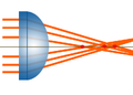
Schematic of spherical aberration - three rays produce three different foci (indicated by a red dot).
Scheme of chromatic aberration - a) simple lens: rays of different wavelengths create three different foci, b) diplet lens: rays of different wavelengths meet at a common focus.
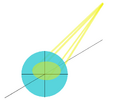
Scheme of coma/astigmatism - light rays do not point parallel to the optical axis, thus creating an asymmetric image (yellow ellipse) in the projection plane (blue circle).


Eyepiece optical system [10][edit | edit source]
The Eyepieces are the last part of the microscope through which the light rays from the light source pass. They participate in the final adjustments of the image. Similar to lenses, they consist of several lenses that work together as a connecting lens; but the number of lenses in the eyepiece is significantly smaller. The eyepiece lenses collectively produce an apparent, magnified, non-inverted image; the overall image produced by the microscope is therefore apparent, magnified and inverted.

In addition to lenses, eyepieces also have an eyepiece diaphragm. According to the position of the eyepiece diaphragm in relation to the lenses, we distinguish two basic design types of eyepieces: the positive eyepiece, in which the diaphragm is located in front of the lenses (closest to the object plane), and the negative eyepiece, where the diaphragm is usually located behind the first lens. The simplest type of positive eyepiece with two lenses is called a Ramsden piece; the simplest type of negative eyepiece is called a Huygens eyepiece.
Similar to lenses, a larger number of lenses will provide better correction of optical defects; so-called plan eyepieces provide the best correction of optical defects.

Basic eyepiece parameters[edit | edit source]
- Focal length of the eyepiece, determined by the distance of the object focal point of the eyepiece from its object principal plane; the focal length determines the magnification of the eyepiece;
- magnification of the eyepiece (see Magnification of the microscope), which is determined by the focal length of the eyepiece; it is essential to determine the total magnification of the microscope, which is the product of the transverse magnification of the objective and the angular magnification of the eyepiece;
- correction of optical defects, which works on a similar principle to the lens − the eyepiece does not have the ability to correct optical defects created in the lens (therefore it is more essential to own a quality lens); the inherent optical defects of the eyepiece are usually eliminated by creating the same optical defect with the opposite effect: for example, the chromatic aberration of the objective is eliminated by using a special doublet of lenses, in which the first lens transmits violet light at a more obtuse angle and the second lens at a sharper angle than red light.
When choosing an eyepiece, it is essential to choose an eyepiece that best matches the given lens - the main factors are the brightness , focal length and numerical aperture of the lens.
Mechanical system[edit | edit source]

In practice, the mechanical system is the system with which the user comes into contact the most. A properly designed mechanical system is a necessary condition for a high-quality microscope: it ensures firm anchoring of the lenses and apertures, the correct angle of the light rays and the object plane, and a very fine mutual displacement of the optical systems and the object plane ("focusing").
The components of the mechanical system are:
- a table with a specimen, through which the object plane passes,
- cross shift , which allows moving the specimen in the subject plane (in practice, it allows the entire specimen to be overlooked),
- macroscrew and microscrew, which allow coarse and finer moving of the stage up and down - by changing the distance between the object plane and the objective, they enable focusing on the given structure,
- revolver, allowing a change of objective (usually a change of objective to an objective with different magnification) so that the distance between the object plane and the objective is maintained,
- tube, separating the objective from the eyepiece, which prevents the penetration of unwanted light rays from the external environment,
- eyepiece focusing ring, which occurs only when the microscope has two eyepieces, one for each eye: in that case, the focusing ring is located on only one eyepiece and allows the correction of the difference between the defects of the eyes by moving the optical system of one eyepiece relative to the optical system of the objective,
- rubber eyecup of the eyepiece, which serves to fix the eyes at the right distance from the eyepiece lenses.
Links[edit | edit source]
Related Articles[edit | edit source]
- The principle of imaging with an optical microscope
- Limit of microscope resolution
- Magnification of the microscope
- Microscope depth of field
- Contrast of the microscope image
Reference[edit | edit source]
- ↑ http://micro.magnet.fsu.edu/primer/anatomy/introduction.html
- ↑ http://zeiss-campus.magnet.fsu.edu/tutorials/basics/fielddiaphragm/index.html
- ↑ http://www.microbehunter.com/the-condenser-aperture-diaphragm/
- ↑ http://web.natur.cuni.cz/~parazit/parpages/mikroskopickatechnika/svetelnamikroskopie.htm
- ↑ http://micro.magnet.fsu.edu/primer/anatomy/objectives.html
- ↑ http://micro.magnet.fsu.edu/primer/anatomy/numaperture.html
- ↑ http://www.photographybay.com/2010/01/02/photography-basics-lens-speed-and-aperture/
- ↑ http://micro.magnet.fsu.edu/primer/anatomy/aberrationhome.html
- ↑ http://micro.magnet.fsu.edu/primer/anatomy/oculars.html
References[edit | edit source]
- NAVRÁTIL, Leoš – ROSINA, Jozef. Medicínská biofyzika. 1 (dotisk 2013) edition. 2005. 524 pp. ISBN 978-80-247-1152-2.
- LEPIL, Oldřich. Fyzika pro gymnázia : mechanické kmitání a vlnění. 3. edition. Prometheus, 2008. ISBN 978-80-7196-216-8.
- MICHAEL, W. Davidson. Basic Concepts in Optical Microscopy [online]. Florida - USA, 2015 [cit. 2015-11-25]. Dostupné z: http://micro.magnet.fsu.edu/primer/anatomy/anatomy.html
- http://web.natur.cuni.cz/~parazit/parpages/mikroskopickatechnika/Optical%20microscopy.pdf
- ABRAMOWITZ MORTIMER, Spring Kenneth R. Basic Concepts in Optical Microscopy. Microscopy resource center [online]. 2012 [cit. 2015-11-25]. Dostupné z: https://www.olympus-lifescience.com/en/microscope-resource/
- OLIVER, Kim. Microscopy Basics. Microbehunter microscopy magazine [online]. Germany, 2014 [cit. 2015-11-25]. Dostupné z: http://www.microbehunter.com/category/microscopy-basics/
- SPRING KENNETH R., Komatsu Hiroshi. Basic Concepts and Formulas in Microscopy [online]. 2015 [cit. 2015-11-25]. Dostupné z: https://www.microscopyu.com/articles/formulas
- Carl Zeiss - Education in microscopy and digital imaging [online]. Germany [cit. 2015-11-25]. Dostupné z: http://zeiss-campus.magnet.fsu.edu/tutorials/index.html
Recommended reading[edit | edit source]
- Interaktivní animace funkcí jednotlivých součástí světelného mikroskopu
- Animace: ovládání světelné aparatury transmisního mikroskopu
- Animace: vliv polní clony na výsledný obraz
- Animace: vliv kondenzorové clony na kužel světla
- Animace: vliv kondenzorové clony na numerickou aperturu objektivu
- Animace: vliv kondenzoru na kontrast výsledného obrazu
- Animace: vliv změn numerické apertury
- Animace: sférická aberace
- Animace: chromatická aberace
- Animace: astigmatismus
- Animace: koma
- Animace: vliv zkreslení na výsledný obraz