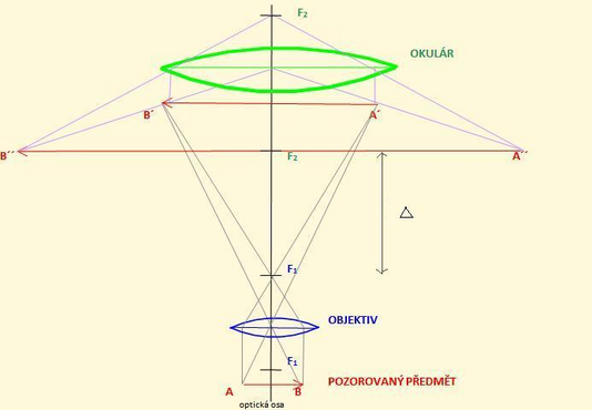
The principle of imaging with an optical microscope
A microscope is an optical instrument that allows you to distinguish structures that are not visible to the naked eye . It includes an optical system , a lighting system and a mechanical device. A microscope increases the angle of view . The visual angle is the angle subtended by the peripheral rays of an object displayed on the retina . As the visual angle increases, the ability to distinguish two points from each other increases. Two points are distinguishable from each other if the rays of these points, which fall on the retinal cells , are at least one retinal cell away from each other.
Optical system[edit | edit source]
The actual display of the object is mediated by the objective and the eyepiece with a common optical axis. The illumination system varies depending on the type of microscope.
Lens[edit | edit source]
It is a connecting system of lenses located closer to the observed object. The lens has a short focal length (1.5-20mm). An object placed just in front of the focal length of the lens (F1) is imaged by the lens to a distance that is greater than twice the focal length of the lens. This image of the object is inverted, real and magnified. The image can be enlarged up to 150×.
Eyepiece[edit | edit source]
It is located closer to the eye. It is a connecting set of lenses that is set up to serve as a magnifying glass . The image formed by the objective is projected between the eyepiece and its focal point (F2). The lens system magnifies this image (up to 20x). The focal length is 10-50 mm. It no longer increases the resolution of the microscope. The final image of the object is magnified, surreal and inverted.
Optical Interval[edit | edit source]
The focus of the objective (F1) and the eyepiece (F2) do not converge, but are far apart. This distance is called the optical interval (Δ). It is around 15-20 cm.
Microscope magnification[edit | edit source]
The total magnification of the microscope is given by the product of the transverse magnification of the objective and the angular magnification of the eyepiece (magnifying glass). The value 250 is the conventional visual distance expressed in mm. Transverse magnification results from the similarity of the triangles. Angular magnification defines the magnification of the angle of view provided by the eyepiece.
Links[edit | edit source]
References[edit | edit source]
- BENEŠ, Jiří, et al. Základy lékařské biofyziky. 1. edition. Praha : Karolinum, 2005. 196 pp. ISBN 80-246-1009-4.
- NAVRÁTIL, Leoš – ROSINA, Jozef, et al. Medicínská biofyzika. 1. edition. Praha : Grada, 2005. 524 pp. ISBN 80-247-1152-4.
- SVOBODA, Emanuel – BARTUŠKA, Karel, et al. Přehled středoškolské fyziky. 4. edition. Praha : Prometheus, 2006. ISBN 80-7196-307-0.


