Radionuclide examinations of the heart
Radionuclide examinations are increasingly used in cardiology , as they can be diagnostic in themselves. We use them to monitor the basic functional and spatial parameters of the heart . The most basic ones include ventriculography, myocardial perfusion scintigraphy, monitoring of metabolic processes and the search for necrosis . Unique to nuclear cardiology is the innervation of the heart .
Myocardial perfusion scintigraphy
This method monitors the accumulation of radiopharmaceuticals in the tissue, which is higher when the monitored area is perfused better. We focus mainly on the left ventricular muscle. It is the most common cardiological examination in the field of nuclear medicine.
The examination can be performed at rest and during exercise, both physical and pharmacological (vasodilators, sympathomimetics). SPECT is used more often to monitor and evaluate the result, planar scintigraphy is also possible, although it has less spatial accuracy.
Indication
Patients with suspected acute coronary syndrome and chronic ischemic heart disease are most often indicated to be monitored for the extent of myocardial blood flow .
Myocardial perfusion scintigraphy plays an important role in the prognosis and further monitoring of myocardial status during and after treatment.
Radiopharmaceuticals
Radiopharmaceuticals with high affinity for myocardial structures are used.
99m Tc-MIBI (methoxyisobutylisonitrile) binds to mitochondria, of which there are many in the myocardium.
201 thallium - a cation, very similar to potassium, its first passage can be monitored, as well as (after 24 hours) the total distribution of potassium (or functional Na / K-ATPase) in the myocardium. The ischemic area shows slower uptake (slow wash-in) and retains the accumulated radiopharmaceutical (slow wash-out) for longer.
Implementation and output
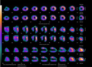
After administration of the radiopharmaceutical, we monitor its accumulation in the heart. Gated scintigraphy can be used for this, which cooperates with the ECG , the recording is the same as for gated ventriculography (Radionuclide examinations of the heart). The ECG gated SPECT, which has a better explanatory value, can also be performed in the same way .
From the ECG gated SPECT we can obtain individual sections of the heart in three planes and 3D reconstruction. This output is excellent for monitoring the placement of a lesion in space. Polar maps compose a 3D image into a two-dimensional "floor plan" by the myocardium, as if we were looking at the heart from the apex. This output is sometimes called the bull's eye . Necrosis then manifests as a failure in a certain part of the circle. This image is usually supplemented by an indication of the course of the coronary arteries and thus finds out in which basin of which artery the ischemia occurred.
In cooperation with computer technology, it is possible to calculate the activity of the radiopharmaceutical in the blood and thus approximately determine the volume of blood flowing through the individual parts. It is also possible to objectively compare the results at rest and under load.
Ventriculography
Ventriculography has the task of showing the heart cavities, their volumes and changes during the heart cycle, or pathology. There are two ways to view the heart cavities:
- first-flow angiocardiography;
- gated ventriculography.
First-flow angiocardiography
A radiopharmaceutical with a short half-life (as many conversions as possible in a short time) is administered as a bolus to a vein as close as possible to the heart (Jugular vein). We then monitor the flow of the radiopharmaceutical through the individual cardiac compartments on a dynamic scintigraphic record. The examination of the heart can be followed by an examination of the blood vessels, but with less accuracy, as the bolus of the radiopharmaceutical is diluted.
Gated ventriculography
Gated ventriculography requires the patient to be connected to an ECG. After iv administration of the radiopharmaceutical, wait a while for the labeled substance to distribute homogeneously in the bloodstream. It is most often labeled with 99m Tc, either autologous erythrocytes or albumin.
The scintillation camera is connected to the ECG. The heart revolution is divided into different numbers (between 16 and 32). Each R oscillation starts a new tracking, the captured images are averaged in individual phases. About 500 heart cycles need to be recorded for the exam to be performed correctly. Examinations can also be performed using the SPECT technique .
Computer technology processes the measured data. The output can then take the form of:
- average heart revolutions - one heartbeat caused by averaging all scintigraphic measurements;
- volume curves - a graph of the volume of cardiac sections, expressed in terms of one averaged cycle, expressed from the activity of the flowing blood.
We can therefore evaluate the volumes of the heart cavities in different phases of the cycle, the rate of their filling, the shape of the cavities and their mutual relations.
Examination of myocardial metabolism
During normal blood circulation, the myocardium consumes mainly fatty acids as an energy source. When the supply of oxygen is reduced (ischemia), there is first a transition to aerobic, later to anaerobic glycolysis. In the phase of anaerobic glycolysis, the myocardial cell no longer contracts, saving energy for its most essential processes. The myocardium is so-called hibernating. When blood flow is restored, the cell gradually returns to its original state and its contractility is restored. If reperfusion does not occur, the cell dies.
Monitoring myocardial viability is of great importance in decision-making in revascularization procedures. A dead myocardium is not worth saving, on the contrary, a hibernating one is.
Viability examination
Radiopharmaceutical 18F-FDG is taken up by the living myocardium as a glucose analogue. By performing PET, it is possible to monitor its distribution and thus very accurately determine the location of viable muscle tissue. Loss of activity means a non-viable myocardium that is unable to accumulate FDG.
Viability can be monitored indirectly by radiopharmaceuticals used in perfusion scintigraphy (thallium, Tc-MIBI) (see above).
Fatty acid metabolism
The uptake of 123 I-labeled fatty acids is not yet widely used in practice. Experimentally, however, they are important in monitoring changes in myocardial metabolic pathways in some diseases.
Search for necrosis
The radiopharmaceutical binds specifically to dead myocardial cells. These sites then show up in scintigraphy as increased accumulation (hot spot ).
Radiopharmaceuticals
99mTc-pyrophosphate binds to the calcium released from the damaged mitochondria of the necrotic muscle.
111In-antimyosin is a monoclonal labeled antibody that is taken up by myosin. It is normally inaccessible to antibodies, it is hidden in the cell. During necrosis and disintegration of the cell membrane, it comes into contact with plasma.
Myocardial innervation
With the help of various radiopharmaceuticals, it is possible to monitor the distribution and functionality of nerve tissue in the heart. We investigate the accumulation of radiopharmaceuticals in synapses and neurons.
We use innervation monitoring to examine transplanted hearts, suspected heart attacks and ischemic heart disease, arrhythmias, heart failure and some neurological diseases.
Links
Related articles
- Recommended examination procedure for suspected acute myocardial infarction
- Heart attack
- Ischemic heart disease
Bibliography
- KUPKA, Karel – KUBINYI, Jozef – ŠÁMAL, Martin, et al. Nukleární medicína. 1. edition. vydavatel, 2007. 185 pp. ISBN 978-80-903584-9-2.

