Coxarthrosis
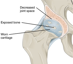
Coxarthrosis can be defined as osteoarthritis of the hip joint. It can affect only one, but also both joints at the same time.
Pathogenesis[edit | edit source]
Factors of heredity and chronic hip overload are involved in the pathogenesis.
Secondary coxarthrosis occurs due to joint incongruence (dysplasia, traumatic changes). It is not the result of aging, although age plays a significant role in its onset. Exceptionally, joint inflammation (specific or non specific) contributes to the development.
The clinical picture[edit | edit source]
Initially exertional hip pain occurs under normal exercise. It also begins to appear at the beginning of the movement. Eventually, it progresses to resting pain, which disrupts normal life (eg sleep).
The range of motion is reduced . It has the greatest effect on the reduction of rotation especially internal rotation. During an objective examination of the joint movement, we therefore find a limitation of the rotations and the pain of the hip joint in the extreme positions. The affected joint tends to occupy the position in which the hip is most relaxed (slight flexion, slight external rotation). In this position, contractures often arise.
If coxarthrosis is caused by dysplasia, a significant difference in limb lengths, a positive Trendelenburg symptom and an atypical position of the affected limb and pelvis may be present .
When walking, there is a typical anti-algae claudication, which is manifested by a quick step through the affected joint.
Arthritic changes are preceded by a state of so-called prearthrosis, which manifests itself in:
- change of joint mechanics (pressure, tension, direction of forces),
- tissue inferiority (congenital, inflammation, injury - also referred to as factor X ).
X-ray image[edit | edit source]
At first, there is a narrowing of the joint space, which is caused by a decrease in cartilage. This is followed by the formation of marginal osteophytes (at the edge of the articular surfaces) and subchondral sklerosis. In the next phase pseudocysts, are formed , which correspond to both parts of the joint ("kissing cysts"). Cystic changes lead to damage to the trophy and thus to the formation of necrotic areas. Pseudocysts collapse over time, causing flattening and deformation of the joint head. The articular cartilage disappears and fibrous, later bony ankylosis develops.
Characteristic senile changes include narrowing of the joint space, loss of bone mass and a slight decrease in the collodiaphyseal angle.
Division[edit | edit source]
Primary coxarthrosis
Secondary coxarthrosis– It develops from a prearthrotic state (it is still reversible). It occurs in the 4th decade and is more common than primary. The most common causes are hip dysplasia, coxitis, osteonecrosis of the head, Perthes' disease, coxa vara, injuries, etc.
According to the affected parts[edit | edit source]
Superolateral form – The most common. Proximolateral migration of the femur with destruction of the head.
Mediocaudal form – Often associated with decreased collodiaphyseal angle and retroversion of the head. Pain during flexion and adduction is typical.
concentric form – Affects the entire hip joint.
All of these forms can be hypertrophic – a marked form of osteophytes or atrophic – loss of bone and cartilage.
Breakdown by severity (of the X-ray image)[edit | edit source]
- Stage I - narrowing of the joint space medially, the beginning of osteophyte formation.
- II. stage - slit narrowing inferomedially, distinct osteophytes, subchondral sclerosis.
- III. stage - slit significantly narrowed, osteophytes, sclerotic changes, cysts of the head and acetabulum, deformation of the shape of the head and acetabulum.
- IV. stage - disappearance of the joint space, sclerosis, cysts, deformity.
Therapy[edit | edit source]
Conservative[edit | edit source]
It consists of a combination of non-pharmacological and pharmacological agents.
Non-pharmacological – Regime measures, weight reduction, rehabilitation, physical therapy, walking with support. Furthermore, the good effects of X-ray therapy, the low dose of which has an anti-inflammatory effect, thereby suppressing reactive synovial inflammation. This therapy is not performed in pregnant women.
Pharmacological – First, we administer analgesics (Template:HVLP) and non-steroidal anti-inflammatory drugs (NSAIDs) (Template:HVLP, Template:HVLP) to relieve intermittent pain. Later, we administer symptomatic slow-acting drugs (SYSADOA - Symptomatic Slow Acting Drugs in Osteo-Arthritis (kys. hyaluronic acidTemplate:HVLP, chondroitin sulfateTemplate:HVLP, glucosamine sulfate), jthe effect of which begins in 3 months, are administered in series.
Operating[edit | edit source]
Osteotomy[edit | edit source]
Change of mutual position and contact of joint surfaces. Less affected areas of cartilage are relocated to more pressure-exposed zones (replacement of devastated cartilage). Valgization osteotomy is most often performed.
Arthroplasty[edit | edit source]
The most common operational solutions. Replacement of destroyed joint socket and head with an endoprosthesis. We have either cemented or uncemented endoprostheses, depending on the method of implantation.
Exceptionally used methods[edit | edit source]
Resection plastic surgery – Removal of the damaged head for about 8 weeks until a fibrous interposition is formed between the proximal end of the femur and the pelvis. We perform it if TEP is not possible and the patient has severe pain. Angulation osteotomy – Extreme solution to a painful condition of the hip. It is performed in severe postdysplastic coxarthrosis. The position of the proximal end of the femur changes, which leads to a change in the load on the affected joint.
Arthrodesis – Joint stiffening in the position of 15 ° flexion and 0-5 ° abduction and neutral rotation. It restores to the patient the possibility of hard physical work while standing, allows full load, but worsens life comfort while sitting.
References[edit | edit source]
Related Articles[edit | edit source]
- Perthes disease
- Osteoarthritis
- Osteoarthritis - surgical treatment
- Osteoarthritis - conservative treatment
References[edit | edit source]
Source[edit | edit source]
- BENEŠ, Jiří. Studijní materiály: Otázky z Ortopedie [online]. [cit. 2011-12-20]. <http://jirben2.chytrak.cz/materialy/orto,trauma_jb.doc>.


