Thromboembolic disease (pediatrics)
Congenital and acquired factors, or a combination of them, can contribute to the development of thrombosis in children. Acquired risk factors are present in 95% of children and 60% of adults. The most common risk for venous thrombosis is an inserted central venous catheter. Thrombosis is not rare in patients with cancer, polytrauma, extensive surgery (CNS), congenital heart defects, systemic autoimmune diseases, especially with the presence of lupus anticoagulant. Hormonal contraception is an important risk factor for teenagers.
For arterial thrombosis, it is catheterization (heart examination, umbilical catheter), the condition after organ transplantation, Kawasaki disease and some congenital heart defects.
Congenital thrombophilic conditions are detected in 10 to 78% of children with thrombosis. These include, for example, resistance to activated protein C (Leiden mutation of the gene for factor V), deficiency of natural protease inhibitors, i.e. AT III, protein C and S, mutation of the gene for prothrombin production, hyperhomocysteinemia. Less common are defects in the fibrinolytic system and dysfibrinogenemia. Heterozygous carriers of these congenital defects rarely develop thrombosis in childhood. As a rule, not only congenital disposition contributes to such thrombosis, but also some of the acquired factors. Homozygous the form of protein C and S deficiency is manifested after birth by purpura fulminans, thrombotic damage to the CNS and sometimes extensive thrombosis of the venous system. In such affected individuals, protein C and S levels tend to be unmeasurable.
The most common locations of venous thrombosis include the area of the inserted venous catheter, as well as the renal and portal veins, the right atrium of the heart, venous shunts, and in adolescents, the venous system of the lower extremities. Arterial thrombosis affects, in addition to catheterized arteries, arteries of transplanted organs, couplings performed in congenital heart defects, CNS arteries, and implanted artificial heart valves.
Clinical picture[edit | edit source]
The clinical symptoms of venous thrombosis are caused by blood stagnation in the affected organ, in the case of arterial thrombosis by organ ischemia.
Laboratory examination[edit | edit source]
Laboratory diagnostics is based on imaging methods: ultrasound with doppler, less often phlebography or arteriography. If pulmonary embolization is suspected, we perform a lung scan, in the case of CNS involvement a CT, MRI or MR angio examination.
Coagulation examination of D-dimers has only supportive value, its negativity testifies against the presence of thrombosis, but does not completely rule it out.
As part of coagulation tests, it is best to take samples before the initiation of anticoagulation therapy, but with LMWH treatment it is possible at the same time. We are investigating:
- aPTT, Quick, INR,
- D-dimers,
- fibrinogen,
- ff. VIII, XI, XII,
- antitrombin III,
- protein C and S, resistance to activated protein C (APC resistance),
- lupus anticoagulant (LA), anticardiolipin antibodies (ACLA),
- KO + diff., biochemistry, liver tests,
- molecular genetics: Leiden mutation, prothrombin mutation (prothrombin 20210),
- homocysteine.
Therapy[edit | edit source]
For treatment in children, we most often use standard heparin or low molecular weight heparin (LMWH) in the acute phase , or thrombolytic drugs. For long-term anticoagulant therapy, oral anticoagulants or antiplatelet drugs (antiaggregants).
Anticoagulation treatment[edit | edit source]
Standard heparin[edit | edit source]
For children, we choose the continuous iv mode. We start with a dose of 75 I.U./kg, we administer this dose over 10 minutes. We continue with a dose of 20 IU/kg/hour. for children > 1 year, for children < 1 year at a dose of 25 IU/kg/hour. The control is performed using aPTT, the recommended range is 60 to 85 s with aPTT norm of 30 to 35 s. This range should correspond to a heparin (anti-factor Xa) level of 0.3 to 0.7 IU/ml. It is an advantage if the laboratory can also examine the heparin level directly. We perform aPTT checks after 4 hours, adjusting the infusion rate according to the table (see below).
If we reach the required range after 4 hours from two subsequent samplings, further checks are carried out after 12 hours, with stable values even once every 24 hours. We also check the blood count daily and the AT III level every other day. When AT III drops < 50%, substitution is in order
| aPTT value | treatment procedure |
|---|---|
| < 50 s | bolus of standard heparin 50 IU/kg + increase infusion rate by 10% |
| 50 to 59 s | increase infusion rate by 10% |
| 60 to 85 s | optimal value |
| 86 to 95 s | reduce infusion rate by 10% |
| 96 to 120 s | interrupt the infusion for 30 min and continue at a rate 10% lower |
| > 120 s | interrupt the infusion for 60 min and continue at a 15% lower rate |
Low molecular weight heparin[edit | edit source]
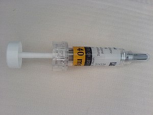
thumb|right|Clexane® Administration of low molecular weight heparin (LMWH, has the same efficacy as standard heparin, but with a lower risk of bleeding. The advantage is that there is less need for laboratory monitoring of the effect. All preparations are administered subcutaneously. Several LMWH preparations (Clivarin®, Clexane®, Fragmin®, Fraxiparine®) are registered on our market. Since their effects are not identical, it is necessary to manage the dosage according to the manufacturer's recommendations.
APTT cannot be used to check efficacy. The effectiveness of LMWH must be verified by determining the anti-factor Xa (aXa) = heparin level. The recommended range is 0.5 to 1.0 aXa IU/ml, a blood sample for control should be taken 3 to 5 hours after administration of the drug.
Oral anticoagulants[edit | edit source]
These preparations reduce the coagulation activity of vitamin K -dependent factors (II, VII, IX, X), but at the same time suppress the activity of K-dependent inhibitors (protein C and S). We currently use warfarin (Warfarin®, Lawarin®). We use PT expressed as INR to check effectiveness(the sensitivity of the reagent used is taken into account in this international standardized ratio). The therapeutic range for children with venous thrombosis is an INR value of 2 to 3, for children with a mechanical heart valve 2.5 to 3.5. Warfarin treatment is started at a dose of 0.15 to 0.40 mg/kg/day, and this dose is continued for 2 to 4 days with daily INR checks. After reaching the desired INR, we reduce the initial dose by approximately 50% and continue with this maintenance dose. Average maintenance doses for infants are 0.3 mg/kg/day, for adolescents 0.1 mg/kg/day. We check the INR every 2 to 3 days, and if we reach the desired range twice in a row, we extend the check intervals to once a week, once every 14 days, etc. The maximum interval between checks should not exceed 4 to 5 weeks.
Warfarin treatment must always be followed by heparin treatment, which is stopped only after reaching an INR value > 2. This is because, in the first days after starting warfarin treatment, K-dependent inhibitors decrease faster than coagulation factors, and we would expose the patient to danger without simultaneous provision of heparin thrombosis (most often it is the so-called warfarin necrosis of the skin). For the same reason, we do not use the recommended initial dose in patients with protein C and S deficiency, but start straight away with the maintenance dose. For regular checks of the effectiveness of the treatment, we follow the scheme in the table (see below).
In case of an overdose of warfarin and simultaneous bleeding manifestations, we apply vitamin K in a dose of 2 to 10 mg after or slowly iv (it must be remembered that after the administration of vitamin K it usually takes a longer time to achieve a sufficient effect of warfarin again; during this time we provide the patient with LMWH) . In case of life-threatening bleeding, we administer frozen plasma, possibly after agreement with the hematologist, prothrombin concentrate.
| INR value | treatment procedure |
|---|---|
| 1,1 – 1,4 | we will increase the dose by 20% |
| 1,5 – 1,9 | we will increase the dose by 10% |
| 2,0 – 3,5 | unchanged |
| 3,5 – 4,0 | we will reduce the dose by 10% |
| > 4,0 | we stop the treatment until it drops < 4.0 and then continue with a 20% smaller dose |
Antiplatelet therapy[edit | edit source]
Acetylsalicylic acid (ASA) is the most commonly used of the preparations that affect the function of blood platelets . It is the drug of choice for some heart diseases, arterial occlusions in the CNS area. The usual therapeutic dose is 3-5 mg/kg/day. It is better to discuss the suitability of other antiplatelet agents (indobufen, dipyridamole, ticlopidine) with a cardiologist, neurologist or hematologist.
Thrombolytic treatment[edit | edit source]
Local thrombolytic treatment[edit | edit source]
Local thrombolytic treatment is mainly used for the patency of catheters. In the case of arterial thrombosis with a serious risk of organ damage or extensive venous thrombosis or pulmonary embolism, we provide general treatment. A few days old thrombus may already be resistant to thrombolysis.
Total thrombolytic therapy[edit | edit source]
Overall thrombolytic treatment always entails a higher risk of bleeding, therefore it is necessary to observe contraindications (bleeding diseases, condition after surgery, trauma, cannulation of a large vessel, ulcer disease, conditions associated with the risk of bleeding into the CNS).
Streptokinase (Streptase®, Kabikinase®) or tissue plasminogen activator (Actilyse®) can be used for thrombolysis . In children, we prefer to use tissue plasminogen activator. The recommended dose is 0.1 – 0.5 mg/kg/hour for a period of 6 hours.
With a good therapeutic effect, the level of fibrinogen decreases, at the same time there is an increase in fibrin cleavage products (D-dimers) and a prolongation of basic coagulation tests (aPTT, PT, TT). Anticoagulant treatment must always be followed after the end of thrombolytic treatment in order to prevent retroembolism of the affected vessel.
Links[edit | edit source]
Source[edit | edit source]
- HAVRÁNEK, Jiří: Trombembolická nemoc. (upraveno)
Related Articles[edit | edit source]
- Krev
- WikiSkripta. Krev - WikiSkripta [online]. [cit. online]. <https://www.wikiskripta.eu/w/Krev>.
- Hemostáza
- WikiSkripta. Hemostáza - WikiSkripta [online]. [cit. online]. <https://www.wikiskripta.eu/w/Hemost%C3%A1za>.
- Koagulace
- WikiSkripta. Hemokoagulace - WikiSkripta [online]. [cit. online]. <https://www.wikiskripta.eu/w/Hemokoagulace>.
Kategorie:Pediatrie Kategorie:Hematologie Kategorie:Vnitřní lékařství

