Pleural effusion

Pleural effusion is defined as the accumulation of more than 10 mL of fluid in the pleural cavity. The free fluid can be of various compositions. One can distinguish exudate, transudate, empyema, hemothorax a chylothorax.
Pathogenesis[edit | edit source]
This depends on the nature of the accumulated fluid.
Transudate[edit | edit source]
.jpg)
Transudate is a protein-poor liquid. It accumulates in cavities when hydrostatic intravascular pressure increases or oncotic pressure decreases (hypoproteinemia). It is usually associated with systemic diseases. The most common causes are congestive heart failure (bilateral), nephrotic syndrome (bilateral), and liver cirrhosis (right).
Exudate[edit | edit source]
It is a protein-rich liquid that accumulates as a result of pathological pleural processes that lead to an increase in pleural permeability or a resorption disorder. It is important to distinguish between exudates associated with an infectious or non-infectious process. Pneumonia is typically associated with infectious diseases. Non-infectious exudates are found in pulmonary embolism, tumor processes (bronchogenic carcinoma, breast cancer, gastric cancer, and malignant lymphoma), and less often in autoimmune systemic inflammation (SLE, rheumatoid arthritis).
Empyema usually arises as a complication of exudate in pneumonia. Hemothorax and chylothorax occur mainly after major trauma.

Bilateral transudate 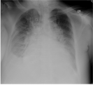
Inflammatory effusion (exudate) 
Hydropneumothorax 
Hemothorax 
Chylothorax


Clinical manifestation and diagnosis[edit | edit source]
The most common manifestation is shortness of breath, which is caused by the compression of the lungs and the restriction of the function of the diaphragm and chest wall. The effusion irritates the pleura, leading to pleural pain and dry irritating cough. In the case of large effusions (e.g., due to a tumor), the mediastinum and venae cavae can become compressed. This causes reduced right ventricular filling and reduced cardiac output.
Physical examination[edit | edit source]
By physical examination, one is able to detect effusions. At the site of effusion, when the accumulated fluid is 300-400 mL, there is weakened or even absent vesicular breathing, dulled percussion, weakened bronchophony, and fremitus pectoralis. When the accumulated fluid is above 400 mL, one can hear weak bronchial breathing.
Imaging methods[edit | edit source]
The most important imaging method is undoubtedly sonography, which detects effusion as low as 50-100 mL. One can also use it to identify fibrin septa and fluid compartments.
A skiagram of the chest reveals effusions that are at least 200-300 mL (blunt costophrenic angle). In bedridden patients, effusions may not be evident until the accumulated fluid becomes 500 mL. In inflammatory effusions, the upper border of the lung has a parabolic shape with a cranially directed convexity. In non-inflammatory effusion, this convexity is smaller.
We use CT a PET/CT to differentiate malignant pleural disorders

.png)
Thoracocentesis[edit | edit source]
It is performed when the underlying cause is not heart failure. Then, the fluid can be analyzed biochemically, cytologically, and microbiologically
- Biochemical examination
- It involves the determination of total protein and LDH to distinguish transudate from exudate (Light's criteria). The examination also detects the presence of amylase (in pancreatitis, esophageal rupture), glucose (rheumatoid effusion, bacterial effusion), and lipids (chylothorax).
- Cytology
- It shows the presence of erythrocytes (in pulmonary embolism), tumor cells, polymorphonuclear cells (in bacterial infections, pulmonary infection), monocytes (in TB, rheumatoid arthritis), or eosinophils (in asbestosis).
- Microbiological examination
- Involves Gram and Ziehl-Neelsen staining, aerobic, anaerobic, fungal, and PCR cultivation.
Biopsy[edit | edit source]
In the case of ambiguity regarding the nature of the effusion, one can perform a skiagram or sonographically guided percutaneous pleural biopsy. If a more precise localization of the origin of the effusion is necessary, one can perform videothoracoscopy with a visually targeted pleural biopsy. This is mainly used for malignancies or tuberculosis of the pleura.
Therapy[edit | edit source]
Treatment is focused on treating the root cause of pleural effusion, which can be either well-controlled (pneumonia, cardiac failure, pulmonary embolism, liver cirrhosis with portal hypertension, etc.) or less difficult to control (disseminated tumors, asbestosis). If the primary causes cannot be further controlled, the treatment goal becomes reducing clinical manifestations, which involves single or repeated thoracocenteses, or drainage, oxygen substitution, and analgesics (especially opiates).
The most reliable method for preventing excessive production of malignant effusions is pleurodesis with intrapleural application of sclerosing agents (talc, bleomycin) through the chest drain. A more effective but invasive method is surgery in the form of simple abrasion of the pleura with subsequent application of talc pleurodesis.
References[edit | edit source]
Related articles[edit | edit source]
External links[edit | edit source]
- MAREL, Miloslav. Novinky v diferenciální diagnostice pleurálních výpotků. Zdravotnické noviny [online]. 2007, y. 56, no. 2, p. příloha Lékařské listy 23–29, Available from <https://zdravi.euro.cz/clanek/priloha-lekarske-listy/novinky-v-diferencialni-diagnostice-pleuralnich-vypotku-287450>. ISSN 1214-7664.
References[edit | edit source]
- KLENER, Pavel. Vnitřní lékařství. 4. edition. Praha : Galén, 2011. 1174 pp. ISBN 978-80-7262-705-9.
- PASTOR, Jan. Langenbeck's medical web page [online]. [cit. 2010-10-27]. <https://www.freewebs.com/langenbeck/>.
- ČEŠKA, Richard. Interna. 3. edition. Praha : TRITON, 2020. 964 pp. ISBN 978-80-7553-780-5.