Hypophysis (SFLT)
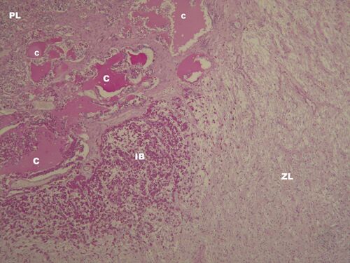
Hypophisis - overview (HE)[edit | edit source]
Description: PL – anterior lobe of hypophisis, ZL – posterior lobe of hypophisis, between there is pars intermedia – in human it's a rudimental middle lobule with Rathke's cleft cysts (C), which are the remnants of Rathke's lumina from pituitary development, C – Rathke's cyst, afunctional, IB – invasion of basophilic cells into the posterior lobe.
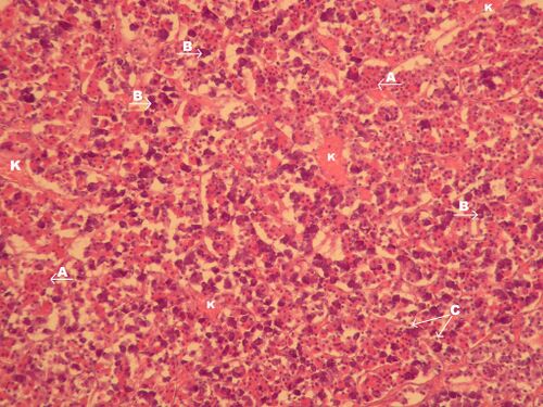
Adenohypophysis - detail (HE)[edit | edit source]
Description: A – acidophil cells, B – basophil cells, C – chromophobe cells K – capillaries with fenestrated endothelium.
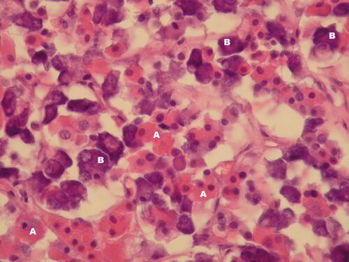
Adenohypophysis – detail (HE)[edit | edit source]
Description: A – acidophil cells, B – basophil cells.
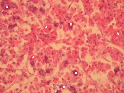
Adenohypophysis – detail (HE)[edit | edit source]
Description: A – acidophil cells, B – basophil cells, C – chromophobe cells.
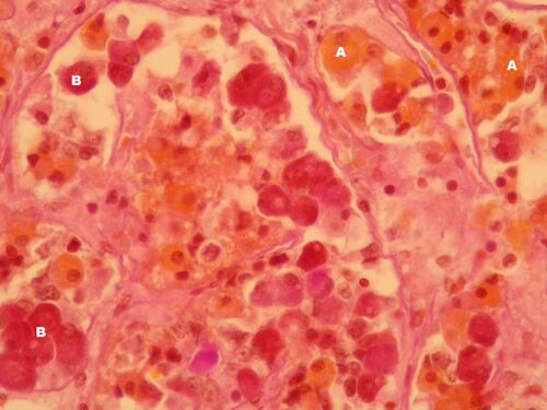
Adenohypophysis – detail (orange G)[edit | edit source]
Description: A – acidophil cells, B – basophil cells
note: in Orange G stain acidophil cells are stained orange and basophil cells (PAS positive) are stained purple.
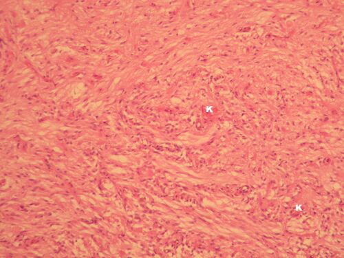
Neurohypophysis - detail (PAS)[edit | edit source]
Description: Neurohypophysis has no epithelial cells, it contains of glial cells (nuclei), neuronal axons from nucleus supraopticus and paraventricularis, synapses and capillaries with fenestrated epithelium (K).
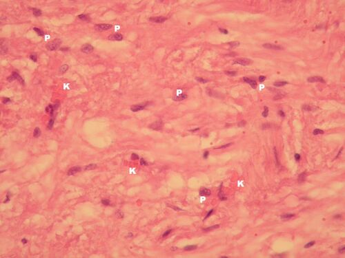
Neurohypophysis – detail (PAS)[edit | edit source]
Description: P – nuclei of the pituicytes (glial cells of the neurohypophysis), K – capillaries with fenestrated endothelium).


