File:CellMembraneDrawing cs.svg
From WikiLectures

Size of this PNG preview of this SVG file: 702 × 371 pixels. Other resolutions: 320 × 169 pixels | 640 × 338 pixels | 1,024 × 541 pixels | 1,280 × 676 pixels | 2,560 × 1,353 pixels.
Original file (SVG file, nominally 702 × 371 pixels, file size: 87 KB)
Summary
| Description |
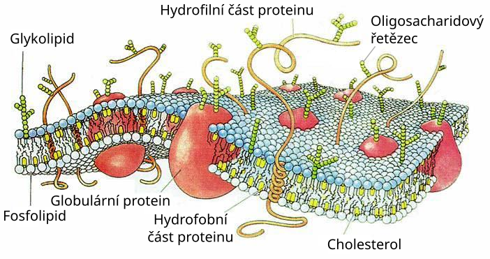
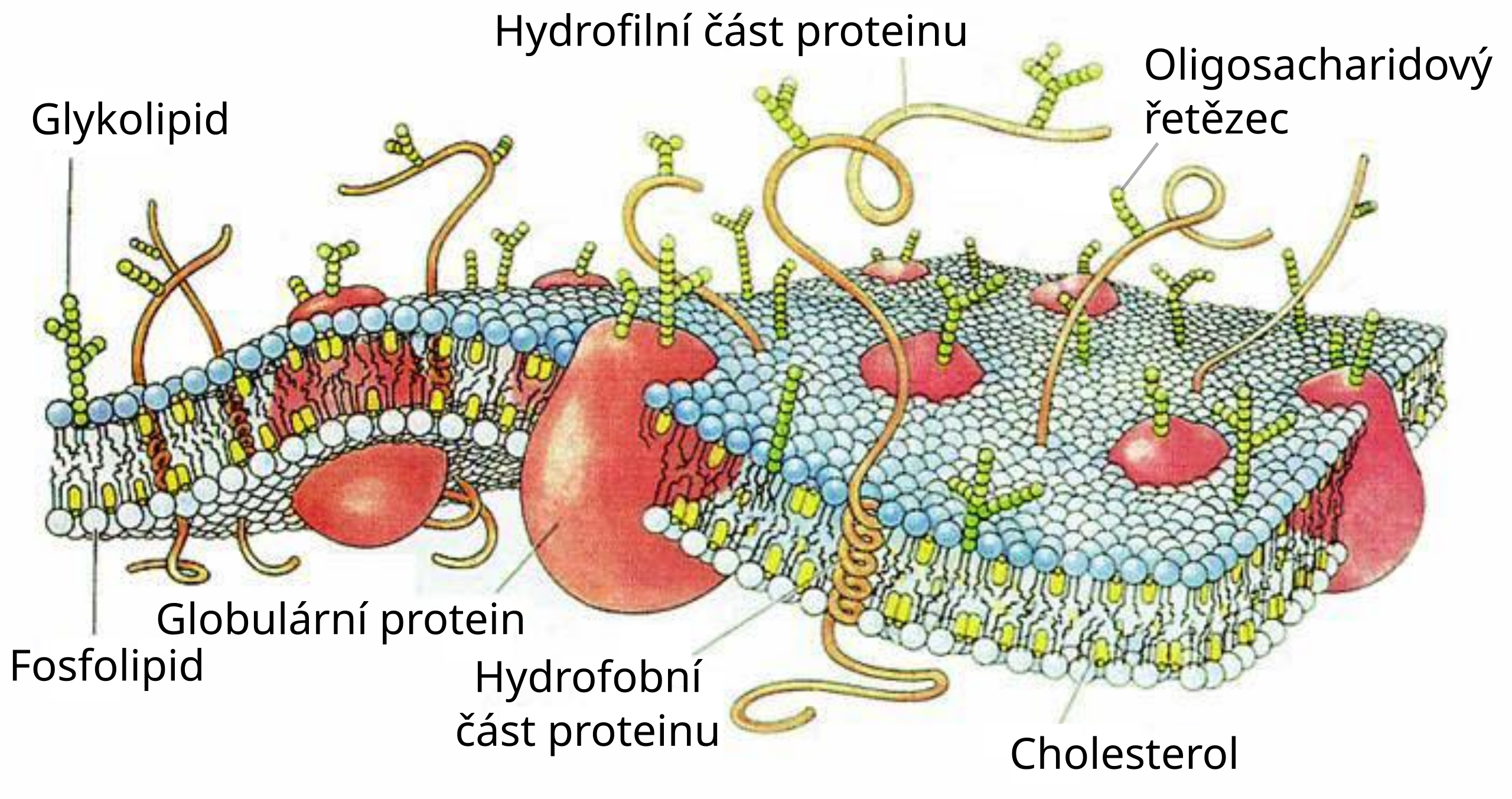
English: Schematic three dimensional cross section of a cell membrane.
There are two major components of this dynamic, fluid, structure: lipids and proteins. A lipid bilayer provides the basic structure within which proteins are free to diffuse. Sugar moieties can be present as part of either proteins (glycoproteins) or lipids (glycolipids). A further important component shown is en:cholesterol; which intercalates between lipid molecules and affects membrane fluidity/stability. Essential Biological Functions: *Immune response *Cell metabolism *Neurotransmission *Photosynthesis *Cell adherence *Cell growth and differentiation Potential Commercial Applications *Drug response monitoring *Chemical manufacturing *Biosensing *Energy conversion *Tissue engineering Source: NIST: These World Wide Web pages are provided as a public service by the National Institute of Standards and Technology (NIST). With the exception of material marked as copyrighted, information presented on these pages is considered public information and may be distributed or copied. Use of appropriate byline/photo/image credits is requested. The drawing was made by Dana Burns, and can also be found in Scientific American, 1985, 253(4), pages 86-90, in the article The molecules of the cell membrane by M.S. Bretscher. |
|||
| Date | (UTC) | |||
| Source | ||||
| Author |
|
|||
| Other versions |
|


| This is a retouched picture, which means that it has been digitally altered from its original version. Modifications: Czech translation. The original can be viewed here: CellMembraneDrawing.jpg:
|
Licensing

|
The copyright holder of this file allows anyone to use it for any purpose, provided that the copyright holder is properly attributed. Redistribution, derivative work, commercial use, and all other use is permitted. |
|
|
Original upload log
This image is a derivative work of the following images:
- File:CellMembraneDrawing.jpg licensed with Attribution
- 2005-07-25T06:29:07Z Matanya (usurped) 702x371 (61721 Bytes) from en wikipedia. schematic three dimensional cross section of a cell membrane. There are two major components of this dynamic, fluid, structure: lipids and proteins. A [[Bilayer|lipid bilayer]] provides the basic structure
Uploaded with derivativeFX
File history
Click on a date/time to view the file as it appeared at that time.
| Date/Time | Thumbnail | Dimensions | User | Comment | |
|---|---|---|---|---|---|
| current | 17:19, 7 July 2011 | | 702 × 371 (87 KB) | wikimediacommons>Icewalker cs | {{Information |Description=Schematic three dimensional cross section of a cell membrane. There are two major components of this dynamic, fluid, structure: lipids and proteins. A lipid bilayer provides the basic structure within which |
File usage
The following 2 pages use this file: