Extraocular muscles
From WikiLectures
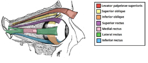
There are 7 extra-ocular muscles - 6 of each responsible for moving the eye ball and 1 is responsible for raising the eyelids.
The extra-ocular muscles are:
Levator palpebrae Superioris (most superior extra-ocular muscle)
- Origin: lesser wing of sphenoid bone (superior-anterior to optic canal)
- Insertion: superior tarsus of the eyelid
- Function: raising the eyelid
- Innervation: superior division of the oculomotor nerve (CN 3).
- Unique feature: some of its inferior fibers are smooth muscle fibers - help with eyelid elevation. Innervated by post-ganglionic sympathetic fibers.
Rectus Muscles (superior, medial and lateral)
Origin: the common tendinous ring (all of the muscles)
Insertion: sclera (posterior to corneoscleral junction)
Function: the axis of the orbit and the axis of the eyeball aren’t the same
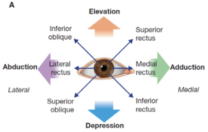
- Superior rectus: moves the eyeball superiorly and medially.
- Inferior rectus: moves the eyeball inferiorly and medially
- Lateral rectus: abduction of eyeball
- Medial rectus: adduction of eyeball
Innervation:
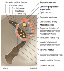
- All except lateral rectus: oculomotor n. (CN 3)
- Lateral rectus: abducent nerve (CN 6)
Superior oblique: medial to the levator palpebrae superioris
- Origin: body of the sphenoid bone.
- Insertion: tendon pass through trochlea insert in sclera (deep to superior rectus).
- Function: abducts, depresses and rotates eyeball medially.
- Innervation: trochlear nerve (CN 4).
Inferior oblique
- Origin: anterior floor of the orbit (maxilla).
- Insertion: Runs posterolaterally crossing the inferior rectus insert in sclera (deep to lateral rectus)
- Function: abducts, elevates and rotated eyeball laterally
- Innervation: oculomotor nerve (CN 3)


