Ear
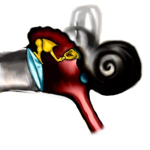
The ear is a sense organ, it is the seat of hearing and balance. It consists of three parts: the outer , middle and inner ear.
External ear[edit | edit source]
Its role is to capture the sound signal and bring it to the drum. It consists of the pinna, the external auditory canal and the eardrum.
The pinna (pinna, auricula) is formed by a plate of elastic cartilage, which is covered by skin.
External ear canal (external acoustic pathway)
- a curved canal that connects the external environment to the temporal bone. When examining the eardrum, it is necessary to straighten this curvature by pulling the pinna upwards and backwards. The eardrum is the medial border of the ear canal.
- it is lined with an epithelial covering passing from the skin (stratified squamous).
- hair follicles are located in the submucosal layer. Hairs - tragi - grow out of them.
- in the submucosa we find sebaceous and modified sweat glands.
- earwax, cerumen, is not produced by sebaceous glands, but by modified sweat glands - glandulae ceruminosae.
The eardrum (membrana tympani) forms the boundary between the outer and middle ear. From the outside, it is covered by a thinned epidermis, and from the inside, it is lined by a single-layer cubic epithelium. Between these layers is a tough connective tissue. The ligament is thinned in the anterior upper quadrant. This part is called the Schrapnell membrane.
Middle ear[edit | edit source]
The tympanic cavity is an irregularly arched space in the middle of the temporal bone. It is lined with a single layer of squamous epithelium.
The middle ear cavity is adjacent to its six walls with different spaces:
- laterally with the tympanic membrane,
- medially with the inner ear,
- dorsally with processus mastoideus,
- ventrally with canalis caroticus,
- cranially with the middle cranial fossa through the tegmen tympani,
- caudally with the fossa jugularis.
The middle ear cavity contains three auditory ossicles: the malleus, the anvil, and the stirrup. They are connected to each other and the hammer is clamped in the drum and the stirrup in the oval window in the medial wall. There are also two muscles: the stapedius muscle and the tensor tympani muscle. It protects the ear from damage by high-intensity sounds - by contraction it dampens the intensity of the transmitted vibrations.
Inner ear[edit | edit source]
It consists of two labyrinths - bony and membranous.
The bony labyrinth consists of a central cavity (vestibulum), three semicircular passages and a cochlea. The sacculus and utriculus are located in the central cavity, the semicircular canals are in the semicircular corridors, and the cochlea is the ductus cochlearis. The snail coils around the modiolus. The modiolus is a bony structure, it has the shape of a cone and the branches of the eighth cranial nerve (ganglion spiral) pass through it. The vestibule and semicircular canals are lined by fibrous cells, thereby forming the mesothelium. The trabeculae depart from the mesothelium, forming the suspension apparatus of the membranous labyrinth. The bony labyrinth is filled with perilymph.
The membranous labyrinth is a system of cavities that evolved from the auditory sacs. This means that it is of ectoderm origin. The sacculus and utriculus developed from the auditory sacs. Semicircular canals are then formed from the saccule and the ductus cochlearis from the utricle. The membranous labyrinth is filled with endolymph.
Sacculus and utriculus [ edit | edit source ]
They are pouches consisting of a fibrous capsule and a single-layered squamous epithelium. There are macules in their wall . They are islands of differentiated sensory cells that are innervated by the vestibular nerve . The receptor elements are called hair cells . Each has on its surface several tens of immobile stereocilia, in the middle of which there is a kinocilia. Between the hair cells are supporting cells that are cylindrical in shape. The layer of cells is covered by a gelatinous layer of glycoprotein . This is probably formed by supporting cells. On the surface of the gelatinous layer there is a layer of crystals, they are called otoliths or statoconia.


