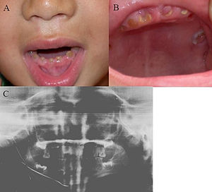
Congenital dental defects
From WikiLectures
Hereditary dentin diseases are divided into dentinogenesis imperfecta (3 types) and dentin dysplasia (2 types). Odontodysplasia can also be included in the genetically determined disorders of dentin.
Dentinogenesis imperfecta[edit | edit source]
- Autosomal dominant hereditary disease;
- dental tissues of mesodermal origin, dentin, pulp, periodontium and cementum are affected ;
- enamel is thin and dentin is fibrous with few tubules;
- we observe other coloring (yellow, brown, gray) and opalescence of the teeth;
- the teeth have less mechanical resistance with a tendency to abrasion.
1st type of dentinogenesis imperfecta
- In patients with osteogenesis imperfecta;
- it is usually caused by a defect in one of the genes encoding collagen;
- the predentin matrix is affected, leading to the formation of amorphous, opalescent dentin without internal organization;
- the incisors are shovel-shaped, the other teeth are lump-shaped;
- medullary cavities obliterate very quickly, sometimes even before cutting;
- the roots of affected teeth are shortened and fragile;
- the temporary dentition is affected more than the permanent dentition.
2nd type of dentinogenesis imperfecta
- Associated with a disorder of genes involved in the formation of bone tissue and dentin;
- the disease is also referred to as "opalescent" dentin;
- this type affects both dentitions equally.
3rd type of dentinogenesis imperfecta
- Bell-shaped teeth;
- teeth described as shell-like (due to thin layer of dentin seen on X-ray );
- open pulp cavities due to rapid abrasion are common;
- found only in Maryland at Brandywine.
Dentin dysplasia[edit | edit source]
- It affects the development and growth of dentin;
- there are morphological changes of the whole teeth or their parts.
The first type
- Rare, etiology not entirely clear;
- the crown is not affected;
- during an X-ray examination, we find abnormalities in the root dentin;
- the roots are significantly shortened, so-called rootless teeth;
- the teeth are mobile and misaligned in the dental arch.
The second type
- In temporary teeth, the manifestations are similar to dentinogenesis imperfecta (mainly color changes);
- permanent teeth usually have a normal shape and color;
- on the X-ray we observe numerous denticles and abnormalities in the structure of the dentine in the permanent dentition.
Odontodysplasia[edit | edit source]
- The defect is due to a disturbance in the early development of the tooth;
- primarily changes in the development of dentin, later also enamel and cementum;
- we observe a hypoplastic crown, a wide pulp cavity and higher tooth decay;
- the teeth of the same quadrant are affected.
Links[edit | edit source]
References[edit | edit source]
- WEBER, Thomas. Memorix of Dentistry : translation 2nd edition, 279 illustrations. 1. edition. Prague : Grada, 2006. ISBN 80-247-1017-X.
- MÁZANEK, George – URBAN, Francis. Stomatological refresher course. 1. edition. Prague : Grada Publishing a.s, 2003. 456 pp. ISBN 80-7169-824-5.
- LIŠKA, Karel. Orofacial Pathology. 1. edition. Prague : Avicenum, 1983. 159 pp.


