Axial Skeleton

The axial skeleton collectively refers to the skeleton of the spine and chest. The skull is often classified with it , but it is functionally and developmentally different. The axial skeleton is the axis of the body, it forms fixed points for the limbs and protects some internal organs. The axial skeleton is evident in all vertebrates. It is segmented, which is caused by development from somites . The skeleton of the spine is made up of vertebrae, the skeleton of the chest is made up of ribs, thoracic vertebrae and the sternum.
Spine[edit | edit source]

The vertebral column forms the axis of the upright body and occupies approximately 35% of body height. It consists of 33-34 vertebrae.
- 7 cervical – vertebrae cervicales (C 1 – C 7 );
- 12 thoracic – vertebrae thoracicae (Th 1 – Th 12 );
- 5 lumbar – vertebrae lumbales (L 1 – L 5 );
- 5 cross – fused in os sacrum (S 1 – S 5 );
- 4–5 coccyx – fused in os coccygis (Co 1 – Co 4,5 ).
Vertebrae[edit | edit source]

The basic structure of a vertebra consists of three mechanically distinct components: body, arch and processes.
Body[edit | edit source]
The body of the vertebra ( corpus vertebrae ) is the supporting part that lies in front. Cranially and caudally, it ends with an almost straight terminal surface for the intervertebral disc. It has a structure typical of short bones - spongiosa with red bone marrow.
Arc[edit | edit source]

The arc of the vertebra ( arcus vertebrae ) is connected to the body from behind and its function is mechanical protection of the spinal cord . The arch has several components: the pedicle , which attaches the arch to the vertebral body; lamina arcus surrounding the spinal cord; foramen vertebrale , which is the opening between the junction of the arch and the body forming the spinal canal; incisura vertebralis upper and lower, which, after the articulation of the two vertebrae, forms on both sides the foramen intervertebrale for the spinal nerve.
Processes[edit | edit source]
Vertebral processes are attached to the arch. It mainly serves to attach the muscles, thereby contributing to the mobility of the joints. We recognize several types of projections: articular processes ( processus articulares ) ensuring the articulation of adjacent vertebrae, are coated with articular cartilage; transverse processes ( processus transversi ), which are paired and extend from the arch laterally; spinous processes ( processus spinosus ) extending backwards. By pulling on the transverse and spinous processes, the vertebrae tilt and rotate.
Types of vertebrae[edit | edit source]
Vertebrae differ according to individual sections.
Cervical vertebrae[edit | edit source]
Vertebrae cervicales have low bodies, except for the atlas. These are wide and short, their terminal surfaces are oval in the shape of a saddle joint. The vertebral foramen is triangular. The spinous processes are short and bifurcated, C1 does not have a spinous process, C 7 has it transformed into a vertebra prominent (a long club-shaped protrusion that can be felt at the junction of the neck and back). The processus transversi are two lateral bumps ( anterior et posterior ), it is actually a stunted rib. Sulcus nervi spinalis is the furrow for the exit of the spinal nerve. The foramen processus transversi is the opening for the course of aa. vertebralis(C 1 – C 7 ) and v. vertebralis (C 7 ). UC 6 is a larger tuberculum caroticum , during bleeding it can press on the common carotid artery . The smallest is C 3 ,the size increases caudally.
- Atlas - has no body but two arches. It bears the articular surfaces for articulation with the os occipitale.
- Axis – carries the dens axis , which is the original body of the atlas. At its top is the apex dentis , which has its own ossifying nucleus.
Thoracic vertebrae[edit | edit source]
The thoracic vertebrae resemble the most general shape of a vertebra. They have tall bodies, with height increasing caudally. The vertebral foramen is round, the spinous processes bend caudally in Th 1 – Th 7 and fold over each other, in Th 7 – Th 12 they straighten again. Foveae costales are the contact surfaces for the heads of the ribs.
Lumbar vertebrae[edit | edit source]
The lumbar vertebrae (vertebrae lumbales) are the largest, transversely very large. The foramen vertebrale is again triangular. The processus spinosi have square plates that are flattened on the side. The processus costarii , which are rudimentary ribs, are also a difference . At the transition of L 5 in the axis of the sacrum , a kink is visible - the promontorium . C 1 –L 5 (to the promontory ) is a movable part of the spine.
Sacrum[edit | edit source]
Os sacrum is part of the spine and girdle of the lower limb. It consists of 5 cruciate vertebrae, which have secondarily fused into bone. Spinous, articular and transverse processes form the cristae sacrales.
Coccyx[edit | edit source]
Os coccygis has already disappeared arches. It is connected to the os sacrum by a synchondrosis.
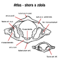
Atlas (top and bottom).
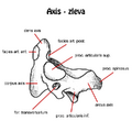
Axis (left).
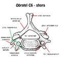
C6 Vertebra (above).
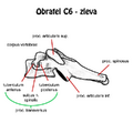
C6 Vertebra (left).
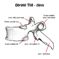
Th8 Vertebra (left).
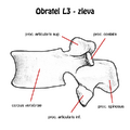
L3 vertebra (left).


Chest[edit | edit source]

The thorax consists of 12 thoracic vertebrae, 12 pairs of ribs and the sternum . The ribs of the first seven pairs reach the sternum and are articulated with it.
Ribs[edit | edit source]
Ribs are long, slender and curved bones. They are attached to the sternum or previous rib by means of cartilago costalis . They have several parts: caput costae (articulated with the vertebra), collum costae , corpus costae and tuberculum costae (articulated with the transverse process of the vertebra, missing on the last two ribs). On the flattened body of the rib, curved and flattened in accordance with the surface of the chest, we distinguish: sulcus costae (recessed inner surface), crista costae (sharp lower edge), angulus costae (stronger curvature of the rib).
We recognize three basic types of ribs.
- The right ribs – costae verae are directly articulated with the sternum (first to seventh pair);
- False ribs - costae spuriae are connected to the previous rib (eighth to tenth pair);
- Free ribs - costae liberae (fluctuantes) end in the abdominal muscles (eleventh to twelfth pair).
The length of the ribs increases from the first to the sixth, then decreases rapidly. The curvature of the rib is three-fold, flat on the circumference of the chest, according to the lower edge (the rib placed on the edge touches the mat only in two places), torsion (the outer surface is vertical at the back, obliquely and upwards at the front). The first, second, eleventh and twelfth ribs differ in shape.
- First rib – costa prima . It is flat and broad with a large tuberculum costae . It has a groove for the sulcus arteriae subclaviae artery of the same name , it is bordered by ridges. It also serves as an attachment for muscles, in front for the scalenus ant. , behind for m. scalenus med .;
- Second rib – costa secunda . It resembles the first rib. Laterally it has tuberositas musculi scaleni post . for the muscle of the same name, continuation of the previous roughness tuberositas musculi serrati ant . serves for one of the teeth of the muscle that is included in the name;
- Eleventh and twelfth ribs – costa undecima et duodecima . They do not have a tuberculum and are only slightly curved.
Sternum[edit | edit source]
The sternum is a flat unpaired bone articulated with the clavicles and with the cranial seven pairs of ribs. It has three main components: the handle of the sternum ( manubrium sterni – cranial part, triangular shape), the body of the sternum ( corpus sterni – the middle, longest part), the sword-like process ( processus xiphoideus – a small process in the caudal part that ossifies during life). It is palpable along the entire length.
- Manubrium sterni - the most prominent formations are the incisura jugularis , which marks the jugular fossa, the paired incisura clavicularis for articulation with the clavicle and the attachment of the cartilages of the first pair of ribs in the incisura costalis prima .
- Corpus sterni – on the cranial edge is the synchondrosa manubriosternalis , which sometimes persists and turns into synostosis at an older age . Between the manubrium and the body of the sternum is the angulus sterni , according to which the other ribs can be counted during the clinical examination, as it articulates with the second rib. Caudally, there are pits for the third to seventh pairs of ribs, on the left they are slightly lower than on the right
- Xiphoideus process – is on the caudal part of the sternum. As a rule, it is pointed, sometimes cross-shaped or with a slit in the middle. The xiphosternal junction is connected in the same way as the manubriosternal junction . The process may remain partially cartilaginous throughout life. Sexual dimorphism is often evident, with females having a shorter process .
Links[edit | edit source]
Related Articles[edit | edit source]
- Spine
- Vertebrae
- Rib Cage
- Ribs
References[edit | edit source]
- ELIŠKOVÁ, Miloslava – NAŇKA, Ondřej. Přehled anatomie. 2. edition. Karolinum;Galén, 2009. ISBN 978-802-4617-176.
- ČIHÁK, Radomír. Anatomie I. 2. edition. Grada, 2001. 516 pp. ISBN 978-80-7169-970-5.