Arteries of the Upper Limb
Arteries[edit | edit source]
Axillary artery[edit | edit source]
The Arteria axillaris is a continuation of the arteria subclavia from the lateral edge of the 1st rib. Next, the axilla runs to the inside of the collum chirurgicum humeri. From there it continues as arteria brachialis.
It supplies the muscles bordering the fossa axillaris, m. infraspinatus, m. deltoideus, parts of the first two intercostals, shoulder joint, skin and part of the mammary gland.[1]
Brachial artery[edit | edit source]
The Arteria brachialis is a continuation of the a. axillaris from the collum chirurgicum humeri. It takes place in the anterior osteofacial space of the arm in the sulcus bicipitalis medialis.
It gives off a strong branch of the a. profunda brachii'', which accompanies the n. radialis in the sulcus nervi radialis, between the heads of the m. triceps brachii. It also gives off the arteria collateralis ulnaris superior' and inferior.
On the front side of the ulnar region in the fossa cubitalis at the level of the collum radii it divides into a. radialis and a.  ;ulnaris. Supplies the arm and elbow joint[2]
Radial artery[edit | edit source]
The Arteria radialis descends from the division of the a. brachialis in the elbow region on the radial side. Her pulse is palpable on the wrist between the brachioradialis and flexor carpi radialis.[1] We find it in the foveola radialis. After crossing the carpus, it runs to the dorsal part of the hand, where between the heads of the ``m. interosseus dorsalis primus it enters the Guiot's space and creates a arcus palmaris profundus.[3]
Gives off the r. palmaris superficialis' contributing to the arcus palmaris superficialis, the branches for the rete carpi palmare and the dorsale and the a . princeps pollicis. It supplies the muscles of the anterior and lateral groups and the skin of the lateral half of the forearm. It is involved in supplying the palm, back of the hand and fingers.[1]
Ulnar artery[edit | edit source]
Arteria ulnaris descends from the division of the a. brachialis under the caput commune ulnare between the m. flexor carpi ulnaris and m. flexor digitorum superficialis.
It gives off a. interossea communis, which subsequently divides into a. interossea anterior, posterior and a. commitans nervi mediani (during the development of the main artery of the limb).[1] It also sends branches for the rete carpi palmare and dorsale, passing through the canalis ulnaris (Guyon's canal)[3]}, issues r. palmaris profundus into arcus palmaris profundus and creates arcus palmaris superficialis .
It supplies the muscles of the anterior and dorsal groups, the m. supinator, the palm, the back of the hand and fingers, the skin in the medial half of the forearm.[1]
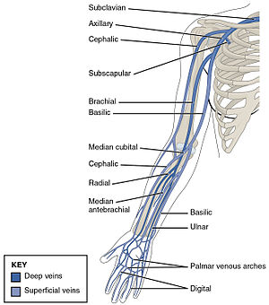
Veins[edit | edit source]
Superficial veins[edit | edit source]
Superficial veins pass from the fingers and palm to the back of the hand. It arises from the rete venosum dorsale manus on the back of the hand and the rete carpi dorsale in the area of the wrist. From the rete venosum dorsa le manus arise v. cephalica and v. basilica, which are used in the place of the cubital fossa for preferential collection of venous blood for laboratory examination.[1]
V. cephalica[edit | edit source]
The Vena cephalica passes to the lateral side of the forearm and after the fascia continues to the elbow region, along the anterolateral side of the arm and in the fossa ovalis infraclavicularis (Mohrenheimi) in the trigonum clavipectorale it enters the axilla and flows into the v.  ;axillaris.[3]
V. basilica[edit | edit source]
The vena basilica passes to the medial side of the forearm, in the lower half of the arm under the superficial fascia through the hiatus basilicus and flows into the vena brachialis.[1]
Deep veins[edit | edit source]
The deep veins are accompanied by the arteries of the same name (up to the axillaris vein, the veins are doubled[3]).
- vv. digitales palmares and vv. metacarpales dorsales
- vv. radiales
- vv. ulnares
- vv. brachials
Links[edit | edit source]
Related Articles[edit | edit source]
References[edit | edit source]
- ↑ a b c d e f g ČIHÁK, Radomír. Anatomy 3. 2. edition. Grada Publishing, 2004. 692 pp. ISBN 978-80-247-1132-4.
- ↑ STANDRING, Susan. Gray's Anatomy : The Anatomical Basis of Clinical Practice. 39. edition. London : Elsevier Ltd, 2005. ISBN 0-443-07168-3.
- ↑ a b c d GIRL, Martin. Limb Anatomy for Winter Autopsy [electronic resource] : (non-saleable script for first-year students of the 3rd LF UK). 1. edition. Charles University, 3rd Faculty of Medicine, 2009. ISBN 978-80-254-3223-5.


