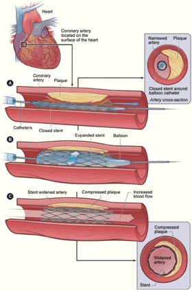
Angioinvasive treatment of arterial blockages and stenoses
|
This article was marked by its author as Under construction, but the last edit is older than 30 days. If you want to edit this page, please try to contact its author first (you fill find him in the history). Watch the discussion as well. If the author will not continue in work, remove the template Last update: Sunday, 18 Dec 2022 at 11.17 pm. |
- Methods of interventional radiology
- PTA (percutaneous transluminal angioplasty);
- placement of stents;
- insertion of Stent grafts or graft-stents;
- local fibrinolysis.
Percutaneous transluminal angioplasty (PTA)
Percutaneous transluminal angioplasty
Percutaneous transluminal angioplasty (PTA) is an invasive therapeutic method in which a special balloon catheter is used to penetrate the lumen of the vessel beyond the stenosis, which is then (mechanically) dilated by inflating the balloon . The dilatation may be followed by the implantation of a stent or stent graft (stenting). Among the causes of stenosis or occlusion of blood vessels are atherosclerosis, fibromuscular dysplasia, conditions after repeated microtraumas, etc.
PTA is used for limb, renal (PTRA, percutaneous transluminal renal angioplasty), coronary (PTCA, percutaneous transluminal coronary angioplasty), supra-aortic arteries, but also for stenoses and vein closures and dialysis AV-shunts.
Indication
Short circular stenoses are generally best suited for PTA .
- Limb arteries – ICHDK grades II.B – IV, improvement of flow before planned bypass , bypass stenoses.
- Supraaortic arteries - manifestations of ischemia of the heart and brain (carotid stenosis, vertebrobasilar insufficiency).
- PTRA – renovascular hypertension based on atherosclerosis or fibromuscular dysplasia.
- PTCA – stable angina pectoris unresponsive to treatment, unstable angina pectoris, AMI , stenosis of the aortocoronary bypass.
- Veins – dialysis shunt stenosis, superior vena cava syndrome .
Contraindication
We distinguish absolute and relative contraindications :
- absolute – unstable condition of the patient, hemodynamically insignificant stenosis, bleeding;
- relative - too long stenosis.
Technique
- Examination of coagulation parameters (INR, APTT, platelet count).
- Before the procedure, we will administer an antiplatelet agent (acetylsalicylic acid , Anopyrin ) and a calcium channel blocker (nifedipine – prevention of vasospasms).
- Prevention of anaphylactic shock in case of allergy to contrast material (antihistamines, corticoids).
- Puncture of a vessel (mostly a. femoralis).
- Heparinization .
- Visualization of the given section of the vascular bed, introduction of the wire into the site of stenosis, and we introduce the catheter along the wire.
- Insufflation of the balloon, dilation of the lesion (keep the balloon inflated for 1-2 minutes).
- Control angiography .
- After heparinization sc two days, at least six months after anti- aggregation (acetylsalicylic acid, Anopyrin®).
Complication
- General (contrast administration) – anaphylactic reaction, renal failure.
- At the puncture site - hematoma, pseudoaneurysm, arteriovenous fistula.
- At the PTA site – dissection, spasm, peripheral embolization, rupture of the artery is a very rare complication.
Stents
- A stent is a reinforcement of a tubular organ, its task is to maintain the lumen and patency of a tubular structure that is narrowed or closed.
Indication
- PTA failure (restenosis, dissection...);
- primary stent implantation – pelvic, coronary, renal artery and internal carotid occlusions that are contraindicated for surgical treatment.
Contraindication
- hypercoagulable conditions (risk of stent thrombosis);
- extreme coiling of the access vascular bed or bed at the site of stent implantation.
- Stents are divided into self- expandable and balloon-expandable .
- After the procedure, patients are heparinized and given antiplatelet agents.
- Complications – as with PTA, in addition, possible stent thrombosis, its migratio,Intimal hyperplasia (stenosis of the stent), compared to PTA, they have a lower risk of restenosis, financially they are comparable to bypassy (but long-term patency is better with bypasses).
Stent-grafts
A stent graft is an endovascular prosthesis, a combination of a stent and a synthetic vascular prosthesis, which is introduced via an endoluminal route, in contrast to classical vascular prostheses, whose implantation is surgical.
- Form
- Grafted-stent – the entire reinforcement consists of a stent that is coated with an artificial material;
- stented-graft – the stent reinforces only the ends of the endovascular prosthesis.
- Indication
- aneurysms ;
- pseudoaneurysms;
- dissecting aneurysms;
- arterial and venous ruptures;
- AV fistula.
Local fibrinolysis
- Local fibrinolysis is indicated for relatively fresh arterial and venous thromboses (including closures of bypasses, AV dialysis shunts, complications of angiography and PTA).
- It consists in the application of a fibrinolytic agent (streptokinase, urokinase, tPA) to the site of thrombosis.
- Contraindication:
- hemorrhagic diathesis;
- kritická končetinová ischemie (it is not possible to wait several hours for fibrinolysis to take effect);
- akutní gastroduodenální vřed;
- conditions after surgery, childbirth or abortion CMP, sepse, malignant tumors.
- In AMI, fibrinolysis can be performed if no more than three hours have passed since the onset of symptoms.
- Execution
- A catheter with end (continuous thrombolysis) or side (pulse thrombolysis) openings is introduced into the thrombus and a fibrinolytic agent is administered with repeated angiographic controls.
- Thrombolysis is usually followed by PTA or stent implantation, or endarterectomy (platelet thrombosis) or bypass.
- Complication :
- Bleeding, peripheral embolization of dissolved thrombus, allergic reaction to a thrombolytic.
Links
References
- ZEMAN, Miroslav. Speciální chirurgie. 2. edition. Praha : Galén, 2006. 575 pp. ISBN 80-7262-260-9.
Source
- BENEŠ, Jiří. Studijní materiály [online]. ©2007. [cit. 14.5.2010]. <http://jirben2.chytrak.cz/materialy/chira/cevni.doc>.
External links
Stent insertion on video: Coronary Angioplasty Stent Placement


