Acute appendicitis

Acute appendicitis is the most common sudden abdominal event , which also occurs at any age. If acute appendicitis is suspected, the patient should always be thoroughly examined and the diagnosis not delayed. Later diagnoses can lead to various complications and serious, life-threatening consequences. Not every appendicitis has a typical course, so it is always necessary to think about it.

Etiology[edit | edit source]
Although the main causes of appendicitis have not been elucidated, the factors that influence the development of this disease are known:
- poor nutrition,
- intestinal contents and the formation of coprolite,
- wedging of a foreign body in the appendix,
- appendix length,
- parasitic diseases, ,
- scar suppression from the outside,
- imbalance of the intestinal flora (especially with more frequent use of antibiotics).
Pathological finding[edit | edit source]
Several types of inflammation can occur in acute appendicitis:
- appendicitis catarrhalis ,
- appendicitis phlegmonosa ,
- appendicitis ulcerosa ,
- appendicitis gangrenosa ,
- perforated appendicitis .
Progression of inflammation may lead to periapendicular infiltrate and diffuse peritonitis.
Clinical picture[edit | edit source]
The classic features of NPB apply to appendicitis :
- Origin from a feeling of full health ,
- sudden start
- abdominal pain
- rapid progression .
The course is largely influenced by the topographic location of the worm-shaped protrusion and its length. In the onset of acute appendicitis, a gradually worsening indefinite and persistent abdominal pain appears , which shifts to the right hypogastrium . In the process, both colic and treacherous pain relief may occur . The pain at McBurney's and Lanz's point is typical .
- McBurney's point:: connection of the navel and spina iliaca anterior superior , at a distance of 2/3 from the navel,
- Lanza's point: the junction of the right and left spina iliaca anterior superior , at a distance of 1/3 from the right spina.
Typical symptoms are nausea and reflex vomiting , loss of appetite and bloating , tachycardia, subfebrile (febrile above 39 ° C rather refutes appendicitis) and Lennander's symptom (difference between axillary temperature and rectal temperature is more than 1 ° C).
Diagnosis[edit | edit source]
Careful history and clinical examination will help determine the diagnosis. With the help of specific symptoms, we can detect irritation of the peritoneum and acute inflammation of the appendix.
- Plenes' sign : percussive pain.
- [Blumberg's sign]]: abdominal pain after releasing pressure on the abdomen, typically at the site of ongoing inflammation, ie in the right lower abdomen in appendicitis.
- Rovsings sign: when the left lower abdomen is pressed, the patient feels pain in the right lower abdomen.
- Psoat symptom: reveals especially the retrocecal localization of the inflamed appendix: during hyperextension or flexion in the hip, the patient feels pain at the appendix site.
- Obturator sign: abduction, flexion and internal rotation cause abdominal pain in pelvic form.
- Döhler's sign - refers to remotely audible, painless, twisting sounds of the cecum during deep palpation in the right lower abdomen later. If the symptom is positive (wriggling is audible and painless), it is most likely acute appendicitis or another inflammatory NPB in the iliac fossa, as the cecum is soft, freely compressible and the perical area is not inflamed nor is there reflex spasm. The sensitivity is close to 100%. Conversely, a negative Döhler's sign (the twisting of the cecum is not audible) does not exclude or confirm acute appendicitis or other NPB.
- 'Procházk's sign - painful blepharospasm during deep painful palpation in the right lower abdomen indicates acute appendicitis (or acute inflammatory affection in the right iliac fossa), the patient does not simulate abdominal pain.
- Paraclinical examination : abdominal ultrasound, event. native image of the abdomen and CT (within diff. dg).
Differential diagnosis[edit | edit source]
When considering the balance, we consider mainly other abdominal events and their typical clinical picture. But we must also keep in mind other abdominal, urological and gynecological diseases.
- Perforated gastroduodenal ulcer (peracute course, history of ulcer disease, free gas in the abdominal cavity).
- Acute gastroenteritis (diarrhoea, history of dietary error, coated tongue, vomiting of undigested food residues).
- Cholecystitis (history of cholecystolithiasis/biliary colic, dietary error, pain radiating below the right costal arch, subicterus and positive Murphy's sign).
- Acute adnexitis (pains are below the groin and symphysis, febrile state).
- Extrauterine pregnancy (symptoms of shock from bleeding, amenorrhoea).
- Burst ovarian cyst, ovarian cyst torsion (women of childbearing age).
- Crohn's disease (long-term diarrhea with colic pains).
- Acetonemic vomiting (in children).
- Pneumococcal peritonitis (in children).
- Pneumonia (contraction of muscles, picture as in peritoneum irritation).
Therapy[edit | edit source]
Acute appendicitis can only be treated surgically by appendectomy , either laparotomically or laparoscopically .
Laparotomy solution[edit | edit source]
We choose pararectal or alternating incision. The advantage of the pararectal incision is that it can be easily enlarged. We choose it, for example, for unclear findings on the abdomen and complicated conditions. For alternating incisions, we penetrate the abdominal wall between mm. obliqui abdominis and m. transversus abdominis , which we lengthwise longitudinally. After opening the peritoneal cavity, we find the cecum and appendix, ligate the vessels (a. Appendicularis) and double-ligate the base of the appendix. The stump is treated with a seroserous suture.
Galerie[edit | edit source]
- Appendix vermiformis.jpeg
Appendix vermiformis
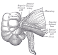
Topography of the appendix

Appendix with cataral inflamation
McBurney's point lies at the junction of the navel and spina iliaca anterior superior, at a distance of 2/3 from the navel.

The Lanz point lies at the junction of the right and left spina iliaca anterior superior, at a distance of 1/3 from the right spina.

CT of the abdomen, arrow shows coprolite, which causes acute appendicitis.

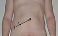
Open appendectomy
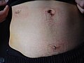
Stitches after laparoscopic appendectomy show, among other things, the previous location of trocars during laparoscopy.


Links[edit | edit source]
Related Articles[edit | edit source]
- Ulcerophlegmonous appendicitis (slide)
- Acute abdomen
- Acute abdomen in children
- Acute abdomen in gynecology
- Differential diagnosis of inflammatory and ileous NPB
- Appendicitis in pregnancy
References[edit | edit source]
- ZEMAN, Miroslav. Special Surgery. 2. edition. Prague : Galen, 2006. pp. 575. ISBN 80-7262-260-9.
- FERKO, Alexander. Surgery in a nutshell : selected chapters. 1. edition. Prague : Grada, 2002. ISBN 80-247-0230-4.