File list
From WikiLectures
This special page shows all uploaded files.
| Date | Name | Thumbnail | Size | User | Description |
|---|---|---|---|---|---|
| 20:19, 21 April 2024 | Thoracic Aorta branches.png (file) | 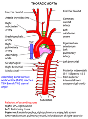 |
85 KB | EshaanM | |
| 19:58, 21 April 2024 | Syntopy of Aortic arch.jpg (file) | 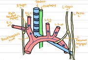 |
241 KB | EshaanM | |
| 23:57, 11 April 2024 | Blastocyst Implantation.png (file) |  |
75 KB | Arya Pawar | |
| 23:49, 11 April 2024 | Development of Blastocyst.png (file) |  |
257 KB | Arya Pawar | |
| 16:35, 6 April 2024 | Classification according number of layers.jpg (file) |  |
45 KB | Janatykondos | |
| 16:31, 6 April 2024 | Basal Lamina.gif (file) | 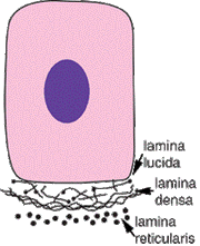 |
12 KB | Janatykondos | |
| 16:10, 6 April 2024 | Cell cycle.gif (file) | 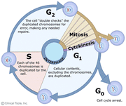 |
33 KB | Janatykondos | |
| 16:05, 6 April 2024 | Meiosis.jpg (file) | 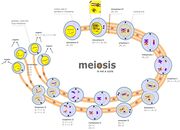 |
156 KB | Janatykondos | |
| 16:03, 6 April 2024 | Mitosis.jpg (file) |  |
32 KB | Janatykondos | |
| 15:28, 6 April 2024 | Immunochemistry.png (file) | 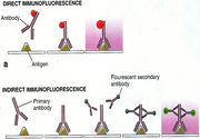 |
253 KB | Janatykondos | |
| 15:26, 6 April 2024 | Enzyme.png (file) | 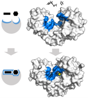 |
54 KB | Janatykondos | |
| 15:15, 6 April 2024 | Cell.jpg (file) | 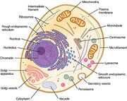 |
111 KB | Janatykondos | |
| 15:12, 6 April 2024 | Cell Surface Specialization.gif (file) | 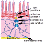 |
18 KB | Janatykondos | |
| 15:09, 6 April 2024 | Cell Membrane 3.png (file) |  |
37 KB | Janatykondos | |
| 15:08, 6 April 2024 | Cell Membrane 2.png (file) |  |
88 KB | Janatykondos | |
| 15:07, 6 April 2024 | Cell Membrane 1.png (file) | 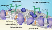 |
269 KB | Janatykondos | |
| 14:56, 6 April 2024 | Gogli apparatus.jpg (file) | 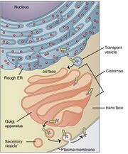 |
84 KB | Janatykondos | |
| 14:55, 6 April 2024 | Mitochondria.jpg (file) | 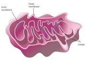 |
26 KB | Janatykondos | |
| 14:46, 6 April 2024 | Picture 2.gif (file) |  |
28 KB | Janatykondos | |
| 13:40, 6 April 2024 | Staining.jpeg (file) | 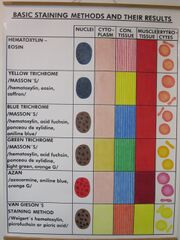 |
239 KB | Janatykondos | {{File |description = Write a description here |author = Author's name and surname |source = Source web address or "own work" |date = Date }} |
| 13:33, 6 April 2024 | Basic Staining.jpg (file) | 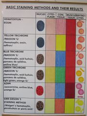 |
239 KB | Janatykondos | |
| 11:18, 29 March 2024 | Syntopy female.png (file) | 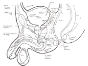 |
265 KB | Nandini | |
| 11:17, 29 March 2024 | Syntopy.png (file) | 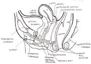 |
346 KB | Nandini | |
| 11:15, 29 March 2024 | Fixation of uterus.png (file) | 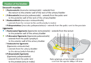 |
302 KB | Nandini | |
| 09:54, 29 March 2024 | Porto caval anastomosis.png (file) | 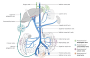 |
531 KB | Nandini | |
| 09:40, 29 March 2024 | 2 stage selection.png (file) | 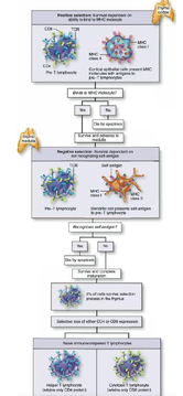 |
166 KB | Nandini | |
| 09:40, 29 March 2024 | Microscope thymus.png (file) | 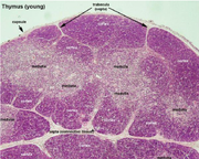 |
749 KB | Nandini | |
| 09:39, 29 March 2024 | Lymphoid organs.png (file) | 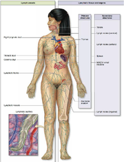 |
379 KB | Nandini | |
| 22:38, 28 March 2024 | Lesser sac.png (file) | 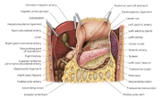 |
160 KB | Nandini | |
| 22:32, 28 March 2024 | Nasal cavity 1.png (file) |  |
98 KB | Nandini | |
| 22:32, 28 March 2024 | Nasal cavity.png (file) | 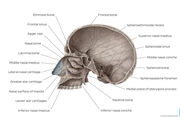 |
93 KB | Nandini | |
| 22:18, 28 March 2024 | Section through ventricles.png (file) | 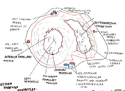 |
254 KB | Nandini | |
| 20:06, 27 March 2024 | Sagittal cross section lower abdominal wall.jpg (file) | 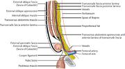 |
50 KB | Shaked.Fru | {{File |description = 1.41Sagittal cross section of the lower abdominal wall at the inguinal region showing anatomic layers at the level of inguinal canal. 1, external oblique fascia (fascia of Gallaudet); 2, external oblique aponeurosis; 3, internal oblique muscle; 4, transversus abdominis muscle; 5, transversalis fascia anterior; 6, external spermatic fascia; 7, Cooper’s ligament; 8, pubic bone; 9, pectineus muscle; 10, transversalis fascia; 11, transversalis fascia posterior lamina; 12, ve... |
| 20:01, 27 March 2024 | Rectus sheath.jpg (file) | 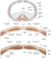 |
27 KB | Shaked.Fru | {{File |description = Relationship between rectus sheath and the parietal peritoneum and transversalis fascia. Section through the abdominal wall superior to the arcuate line. Section through the abdominal wall inferior to the arcuate line. |author = Schuenke M, Schulte E, Schumacher U. THIEME Atlas of Anatomy. General Anatomy and Musculoskeletal System. Illustrations by Voll M and Wesker K. 3rd ed. New York: Thieme Medical Publishers; 2020. |source = : 3.2.2 Rectus Sheath. In: Wike... |
| 19:57, 27 March 2024 | Muscles of abdominal wall.jpg (file) | 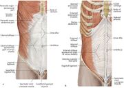 |
39 KB | Shaked.Fru | {{File |description = Muscles of the abdominal wall. Right side, anterior view. Superficial abdominal wall muscles. Removed: external oblique, pectoralis major, and serratus anterior. |author = Schuenke M, Schulte E, Schumacher U. THIEME Atlas of Anatomy. General Anatomy and Musculoskeletal System. Illustrations by Voll M and Wesker K. 3rd ed. New York: Thieme Medical Publishers; 2020. |source = : 3.2.1 Muscles of the Anterolateral Wall. In: Wikenheiser J, Voll M, Wesker K, ed. Clin... |
| 14:08, 16 December 2023 | Ovarium..jpg (file) | 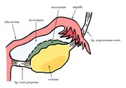 |
341 KB | Viktorie Koberová | |
| 14:07, 16 December 2023 | Pelvic floor.jpg (file) | 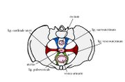 |
316 KB | Viktorie Koberová | |
| 14:06, 16 December 2023 | Uterus..jpg (file) | 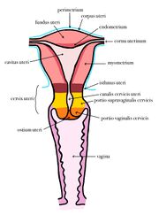 |
370 KB | Viktorie Koberová | |
| 14:03, 16 December 2023 | Stomach..jpg (file) | 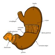 |
311 KB | Viktorie Koberová | |
| 14:02, 16 December 2023 | Stomach – picture.jpg (file) | 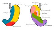 |
338 KB | Viktorie Koberová | |
| 14:01, 16 December 2023 | Stomach – anatomy.jpg (file) | 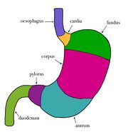 |
171 KB | Viktorie Koberová | |
| 13:44, 16 December 2023 | Basal ganglia.jpg (file) | 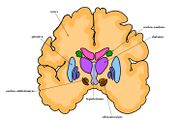 |
475 KB | Viktorie Koberová | |
| 13:40, 16 December 2023 | Tongue.jpg (file) | 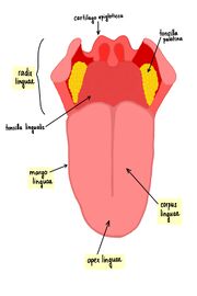 |
357 KB | Viktorie Koberová | |
| 13:38, 16 December 2023 | Kidney – structure.jpg (file) | 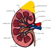 |
540 KB | Viktorie Koberová | |
| 10:48, 16 December 2023 | Brain.jpg (file) | 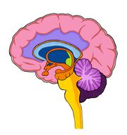 |
413 KB | Viktorie Koberová | |
| 10:43, 16 December 2023 | Ossa carpi.jpg (file) | 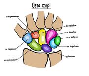 |
326 KB | Viktorie Koberová | |
| 14:24, 9 December 2023 | Koberová fotka.jpg (file) |  |
1.63 MB | Viktorie Koberová | |
| 13:22, 9 December 2023 | Zavedená LM.jpg (file) |  |
598 KB | Cateducated | |
| 17:28, 19 November 2023 | Abductor hallucis.png (file) | 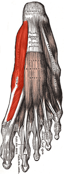 |
164 KB | Darsh jhala | {{File |description = Write a description here |author = Author's name and surname |source = Source web address or "own work" |date = Date }} |
| 11:49, 12 November 2023 | Peroxynitrite ion 2D.png (file) |  |
9 KB | Sam02 |