File:Siphovirus.tiff
From WikiLectures

Size of this preview of this TIF file: 594 × 599 pixels. Other resolutions: 238 × 240 pixels | 476 × 480 pixels | 761 × 768 pixels | 1,048 × 1,057 pixels.
Original file (1,048 × 1,057 pixels, file size: 1.09 MB, MIME type: image/tiff)
Summary
| Description |
English: Electron micrographs of bacteriophages from Cutibacterium acnes (formerly Propionibacterium acnes). Phages were negatively stained with 0.75% uranyl formate and subjected to transmission electron microscopy. The phages have a head of approximately 55 nm in diameter, loaded with genetic material. Their tails have a size of 150 × 10 nm and are flexible and non-contractile. The upper micrographs show Propionibacterium phage PAD40 and PAD11 (NBBI TaxIDs 504513 and 504513 respectively). |
| Date | |
| Source | BMC Microbiology 2008, 8:139 doi:10.1186/1471-2180-8-139 |
| Author | Rolf Lood, Matthias Mörgelin, Anna Holmberg, Magnus Rasmussen and Mattias Collin |
Licensing
This file is licensed under the Creative Commons Attribution 2.5 Generic license.
- You are free:
- to share – to copy, distribute and transmit the work
- to remix – to adapt the work
- Under the following conditions:
- attribution – You must give appropriate credit, provide a link to the license, and indicate if changes were made. You may do so in any reasonable manner, but not in any way that suggests the licensor endorses you or your use.
File history
Click on a date/time to view the file as it appeared at that time.
| Date/Time | Thumbnail | Dimensions | User | Comment | |
|---|---|---|---|---|---|
| current | 16:31, 2 December 2011 | 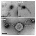 | 1,048 × 1,057 (1.09 MB) | wikimediacommons>Alexbateman |
File usage
The following page uses this file: