Stress-activated channels


What are stress-activated channels?[edit | edit source]
Stress-activated channels are membrane proteins capable of responding to mechanical stimuli. This means they can be activated for example by movement or pressure, which leads e.g. to opening and closing of the channels. Stress-activated channels are also called mechanosensitive (ion) channels (MSCs) and function as mechanotransducers which are capable of generating both electrical and ion flux signals as a response to external or internal stimuli. MCSs are the basis of the senses of hearing and touch and in addition sense the stress needed for muscular coordination.
Where do we find them?[edit | edit source]
Stress-activated ion channels perform important functions in many different areas of our body. For example require pressure-dependent myogenic constriction resistance arteries these channels for regulation in the smooth muscle of the arteries. They have been found to be used for volume sensing in animals and blood pressure regulation. Bacteria have been shown to relieve hydrostatic pressure through those channels. They are also found in the human ear, where they perform the transmission of the soundwave signal into an electric signal which then is lead to the brain. In the inner ear we find the cochlea containing the organ of corti. This organ contains the cells responsible for hearing, the hair cells. The bottom of these cells is attached to the basilar membrane, and the stereocilia (“hairs”) are in contact with the tectorial membrane. Stress-activated channels are present in the membrane of the stereocilia and change the mechanical acoustic wave signal into an impulse/action potential that can then be transmitted to the brain resulting in actual hearing.
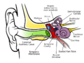
Parts of the ear

How does it work?[edit | edit source]
Outer hair cells have a special function within the cochlea, where sound waves cause the basilar membrane to vibrate up and down, which creates a shearing force between the basilar membrane and the tectorial membrane, causing the stereocilia to bend back and forth. This movement of the cells opens the mechanosensitive potassium channels of the stereocilia causing positively chaged potassium ions to flow into the hair cells. This increases the membrane potential, causing other voltage gated calcium channels in the hair cell membrane to open and letting calcium ions flow into the cell too. The calcium ions are now the trigger causing neurotransmitter release and stimulating the auditory nerve which then sends the sound signals as an action potential to the brain which then can interprete it as e.g. music or speech.
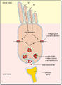
Opening of potassium channels

What makes them so important and where problems can occur?[edit | edit source]
The movement of the haircells and thus the opening of stress-activated potassium channels in our ears provide the converting of a wave signal into an electirc signal that the brain can understand which is why they are one of the most important parts of the sence of hearing. They allow us to hear even very quiet sounds but when they are exposed to loud music or noise, the hair cells can be seriously damaged. Loud sounds are large pressure waves, which can bend the stereocilia too far and permanently damage them. Cochlear hair cells cannot grow back which then leads to a loss of hearing.
References:[edit | edit source]
Baylor College of Medicine, https://www.bcm.edu/healthcare/care-centers/otolaryngology/patient-information/hearing-hair-cells
Leslie Samuel, https://www.youtube.com/watch?v=lDXVZOU_f_E
Leslie Samuel, https://www.youtube.com/watch?v=GpkD8AZTFCs
Brandon Pletsch, https://www.youtube.com/watch?v=46aNGGNPm7s
Animacionesplus (Youtube channel), https://www.youtube.com/watch?v=1JE8WduJKV4