Heart Development
|
This article was marked by its author as Under construction, but the last edit is older than 30 days. If you want to edit this page, please try to contact its author first (you fill find him in the history). Watch the discussion as well. If the author will not continue in work, remove the template Last update: Monday, 06 Jul 2015 at 2.14 am. |
The Heart[edit | edit source]
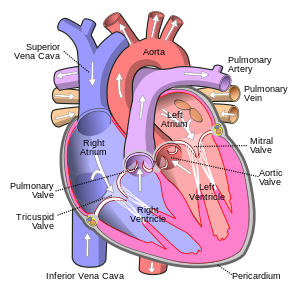
The Heart is a muscular organ that is involved in the pumping of blood around the body and thus bringing about exchange of Oxygen and Co2 between blood and cells; a function important for survival. The heart consists of 4 chambers; two atrium (left and right) and two ventricles (left and right). These chambers are divided by valves and the left side and right side are separated by an inter muscular and inter membranous septum.
Deoxygenated blood enters the Right Atrium, via the Superior Vena Cava, passes into the Right Ventricle and out through the Pulmonary Artery to the lungs where exchange of Oxygen and Co2 will take place, and then blood re-enters the Left Atrium via the Pulmonary Artery as oxygenated blood. This then passes out via the Aorta to the rest of the body.
The Development of the heart is thus an important part of embryological development and occurs in the following way:
3rd week[edit | edit source]
During the 3rd week of Embryological development, blood islands form in the Mesoderm surrounding the embryonic yolk sac. The blood islands then collate and form Hemangioblasts, that are precursors for blood cells and vessels. Angioblastic cords appear in the Cardiogenic Mesoderm and canalize into the heart tube by lateral embryonic folding. The tube then elongates and develops into the Aoritc roots, Bulbous Cordis, Primordial Ventricle, Atrium and Sinus Venosus.
4th week[edit | edit source]
During the 4th week, endocardial cushions are present on the dorsal and ventral wall of the AV canal and these fuse, thus separating the primordial atrium from the primordial ventricle. The Septum Primum grows from the roof of the Primordial Atrium, towards the endocardinal cushions and this division allows formation of the left and right atrium.
7th week[edit | edit source]
The ventricular septum has both an inter muscular and inter membranous one. The inter muscular septum comes about with lying on the floor of the ventricle and then the dilation of ventricles, causing its medial walls to fuse. The inter membranous septum forms during the 7th week of development; a crescent-shaped interventricular foramen that allows communication between right and left ventricles exists before this septum. However after the 7th week, this foramen closes due to the bulbar ridges fusing with the endocardial cushion and so forming an inter-membranous septum.
During fetal development, the pulmonary system of the fetus is non-functional and so the fetus relies on the mother's contribution via the placenta. Thus when blood enters the Right Atrium, it cannot pass into the Pulmonary Artery, as it is non-functional, and so it has to be shunted from the Right Atrium to the Left Atrium so that blood can pass to all the cells in the body. This shunting tends to occur when a foramen primum begins to develop between the free edge of the septum and endocardinal cushion and so blood can pass freely from the Right Atrium to the Left Atrium. This foramen primum then is eventually replaced by a foramen secundum and this develops into the Foramen Ovale.
At birth, with the baby's first breath, this Foramen Ovale tends to seal up, as the pulmonary circulation becomes active and pressure in the Left Atrium increases significantly.
Bibliography[edit | edit source]
Before We Are Born: Essentials of Embryology and Birth Defects, Saunders; 6 edition

