Circulation changes after birth
The prenatal circulation is different from postnatal circulation after the fetus is disconnected with the umbilical vessels.
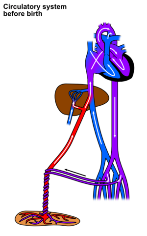
In prenatal circulation, the oxygenated blood comes from umbilical vein. Most of it bypasses the liver by "ductus venosus." Then it goes straight to the right atrium. There is another supply that right atrium gets blood from, which is superior vena cava. Blood from inferior vena cava is much more highly oxygenated, and it goes into the foramen ovale to pass through to the left atrium and left ventricle. Blood coming out of aorta goes mainly to the head. The blood from superior vena cava pass down from right atrium to right ventricle. But since there is high resistance in pulmonary arteries as the lungs are not expanded, the blood is drained into arch of aorta via "ductus arteriosus." Then this mixed blood comes out of fetus by umbilical arteries.
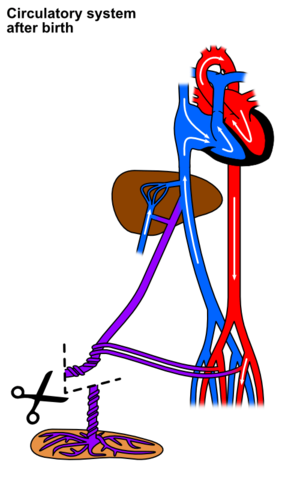
At birth, the umbilical arteries and vein is disconnected. Primary changes n pulmonary and systemic vascular resistances at birth make circulation change. Due to loss of tremendous blood flow through the placenta, the systemic vascular resistance at birth doubles. As resistance rises, aortic pressure increases. Furthur in pressure in left ventricle, left atrium increases as well. Due to expansion of the lungs, the pulmonary vascular resistance greatly decreases. Then the pulmonary arterial pressure is reduced so does pressure in right atrium and right ventricle.
Therefore, blood wants to flow from high left atrial pressure to low right atrial pressure. It is backward through foramen ovale compared to fetal circulation. But there’s a valve lies over foramen ovale on the left side of the atrial septum, so closing of foramen ovale occurs. Now the forame ovale is called "fossa ovalis"
Because of lost connection of umbilical vessels, closure of ductus arteriosus and closure of ductus venosus occur. Closure of ductus arteriosus is by smooth muscle contraction. Smooth muscle on ductus arteriosus occurs to close the blood flow completely in 1-8days. it is further replaced by fibrous tissue, called ligamentum arteriosum. This contraction of smooth muscle occurs becuase of the increase in availability of oxygen. At birth, opposite direction of blood flow from aorta to pulmonary artery supplies more oxyginated blood than before. The partial pressure of oxygen is used to be 15 - 20 mmHg before, but now it is 100mmHg. It is confirmed that the degree of smooth muscle contraction is highly dependant on more availability of oxygen. Closure of ductus venosus is also caused by strong contraction of muscle wall of ductus venosus, but the cause of this contraction is not revealed yet. This structure is changed into fibrous ligamentum venosum. It is sometimes continuous with the round ligament of the liver.
Further changes are shown in the picture. The umbilical vein becomes round ligament of the liver, and the umbilical arteries become medial umbilical folds.
1.Blood volume: immediately after birth averages about 300ml. (75mL additional blood from umbilical cord – if it’s still attached) But the fluid is lost into the neonate’s tissue spaces again - 300mL maintained. 2.Cardiac Output: in average 500ml/min. 3.Arterial Pressure: 70/50, but it increases slowly during next several months to about 90/60
GUYTON, Arthur C. Medical Physiology. 11th edition. 2006. Chapter 83: Fetal and Neonatal Physiology. ISBN 9780721602400.


